- Title
-
Perfluorooctanoic acid disrupts thyroid hormone biosynthesis by altering glycosylation of Na+/I- symporter in larval zebrafish
- Authors
- Cai, Z., Zhou, G., Yu, X., Du, Y., Man, Q., Wang, W.C.
- Source
- Full text @ Ecotoxicol. Environ. Saf.
|
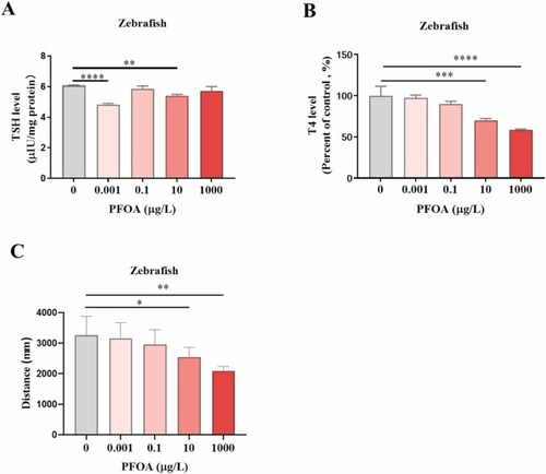
PFOA disrupted thyroid function and locomotor behavior of zebrafish larvae. A Concentrations of TSH in zebrafish larvae were measured by ELISA at 7 dpf. n = 3. B Levels of T4 in zebrafish larvae were measured by ELISA at 7 dpf. n = 3. C The effect of PFOA on locomotor behavior of zebrafish larvae. n = 16. Asterisks indicate statistically significant differences (one-way ANOVA with Tukey's HSD post hoc test,*:p < 0.05; **:p < 0.01, ***:p < 0.001; ****:p < 0.0001). The treated groups were compared with the control group. |
|
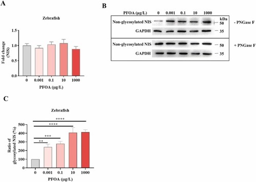
The effect of PFOA on NIS mRNA levels and glycosylation of NIS protein in zebrafish larvae. A The zebrafish eggs were exposed to different PFOA concentrations for 7 days. Then, the mRNA levels were measured by real-time PCR. The treated groups were compared with the control group. n = 3. B zebrafish eggs were treated with different concentrations of PFOA for 7 days. NIS protein levels were analyzed using Western blot. GAPDH was used as a loading control for normalization. C The band intensities were calculated using Image J software. The ratios of glycosylated NIS to non-glycosylated NIS were plotted. one-way ANOVA with Tukey's HSD post hoc test,n = 3,**:p < 0.01, ***:p < 0.001; ****:p < 0.0001). The treated groups were compared with the control group. |
|
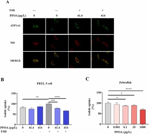
PFOA disrupted membrane expression of NIS protein and inhibited iodine uptake. A Representative confocal immunofluorescence microscopy image of NIS localization in FRTL-5 cells after PFOA exposure for 48 h in the presence of TSH. ATP1A1 (Sodium/potassium-transporting ATPase subunit alpha-1) was used as a cell surface marker. NIS and ATP1A1 were visualized in red and green, respectively. Yellow fluorescence is equivalent to the co-localization of the NIS with ATP1A1. B Effect of PFOA exposure on iodide uptake in FRTL-5 cells. FRTL-5 cells were treated with various PFOA concentrations for 48 h, then the iodide uptake ability of FRTL-5 cells was measured. Treatment group compared with the TSH group. C Effect of PFOA exposure on iodide uptake in vivo. Zebrafish eggs were treated with different PFOA concentrations for 7 days. After treatment, the iodide uptake ability was measured. The treated groups were compared with the control group. 41.4 µg/L (0.1 µM); 414 µg/L (1 µM) One-way ANOVA with Tukey's HSD post hoc test,*:p < 0.05; **:p < 0.01, ***:p < 0.001; ****:p < 0.0001). |
|
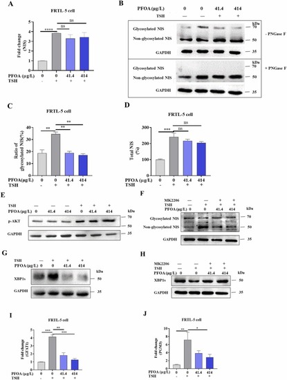
The effect of PFOA on NIS and TSH-dependent signaling in FRTL-5 cells. A FRTL-5 cells were treated with various PFOA concentrations for 48 h, then the NIS mRNA levels were measured. B Cells were treated with different concentrations of PFOA for 48 h. Then, cell lysates were used to measure NIS expression and glycosylation using Western blot. GAPDH was used as a loading control for normalization. The band intensities were calculated using Image J software. The ratios of glycosylated NIS to non-total NIS (C) and the total NIS level (D) were plotted, respectively. PFOA-treated groups compared with the untreated group in the presence of TSH. E Cells were treated with different concentrations of PFOA for 30 min. After treatment, cells were lysed to detect AKT phosphorylation levels using Western blot. GAPDH was used as a loading control for normalization. F Cells were treated with TSH and different concentrations of PFOA in the presence of or in the absence of MK2206 for 48 h. Then, cells were lysed for Western blot analysis to detect NIS glycosylation levels. GAPDH was used as a loading control for normalization. G Cells were treated with different concentrations of PFOA with or without TSH for 48 h. Then, cells were lysed to measure XBP1s protein levels using Western blot. H Cells were treated with TSH and various concentrations of PFOA in the presence of or in the absence of MK2206 for 48 h. Then, cells were lysed to detect NIS glycosylation levels using Western blot. GAPDH was used as a loading control for normalization. I, J Cells were treated with different concentrations of PFOA for 48 h. Then, real-time PCR was performed to detect the transcription levels of GFAT1 (I) and PGM3 (J). 41.4 µg/L ( 0.1 µM); 414 µg/L ( 1 µM). One-way ANOVA with Tukey's HSD post hoc test,n = 3,*:p < 0.05; **:p < 0.01, ***:p < 0.001; ****:p < 0.0001). The treated groups were compared with the control group. |
|
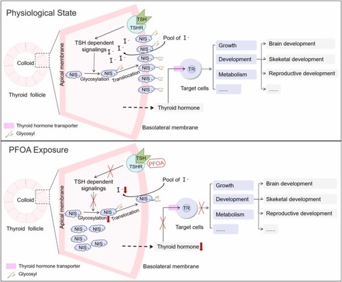
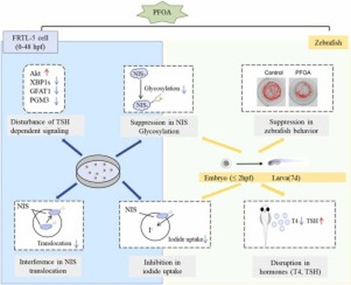
Schematic diagram illustrating the mechanisms by which PFOA exposure disrupts thyroid hormone synthesis. In the physiological state, TSH triggers TSH-dependent signalings to regulate gene expression and protein glycosylation of NIS, which is responsible for iodide uptake and is the critical molecule for thyroid hormone synthesis. Thyroid hormones exert their effects on target cells throughout multiple systems to regulate various physiological processes, including metabolism, growth, and development of the body. Exposure to PFOA can disrupt the glycosylation process of NIS, which in turn inhibits the translocation of NIS to the plasma membrane. This inhibition leads to a decrease in the uptake of iodide and subsequently results in reduced levels of thyroid hormones in zebrafish larvae. TSH: Thyroid Stimulating Hormone; TSHR: Thyroid Stimulating Hormone Receptor; NIS: Na+ /I− Symporter; TR: Thyroid Hormone Receptor; I-: iodide ion; PFOA: Perfluorooctanoic Acid. |
|
|

