- Title
-
Zebrafish PEX1 Is Required for the Generation of GABAergic Neuron in p3 Domain
- Authors
- Ryu, J.H., Kim, M., Kim, A., Ro, H., Kim, S.H., Yeo, S.Y.
- Source
- Full text @ Dev Reprod
|
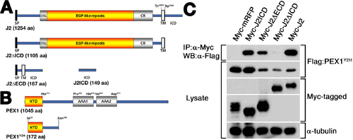
The intercellular domain (ICD) of zebrafish Jagged2 interacts with the N-terminal domain (NTD) of PEX1. (A, B) Schematic diagrams of the constructs used to yeast-two hybrid screening and Western blotting. (A) Schematics of Myc-tagged Jagged2 (J2) constructs. (B) Schematics of PEX1. An identical 516 bp-long fragment of PEX1 called PEX1Y2H isolated from yeast-two hybrid screening using J2ICD as the bait. (C) Co-immunoprecipitation (Co-IP) performed using the lysates of NIH 3T3 cells transfected with expression vectors for Myc-tagged J2 variants and Flag-tagged PEX1Y2H, as indicated. Immunoprecipitated with and anti-Myc antibody and separated by SDS-PAGE followed by transfer to a PVDF membrane and immunoblotting with anti-Flag antibody. Myc-tagged RFP used as a control for Co-IP. Antibody for anti-α-tubulin used as an internal control. SP, signal peptide domain; DSL, Delta/Serrate/LAG-2 domain; CR, cysteine-rich domain; TM, transmembrane domain; AAA, AAA-ATPase domain. |
|
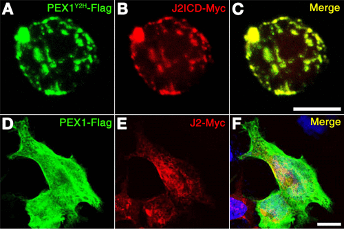
Zebrafish PEX1 partially co-localized with Jagged2 in the plasma membrane. (A–C) Expression vectors for PEX1Y2H and J2ICD were transfected into P19 cells. Confocal images indicated the Flag-tagged PEX1Y2H in the green channel (A) and the Myc-tagged J2ICD in the red channel (B). The merge image is displayed (C). (D–F) Expression vectors for PEX1 and J2 were transfected into NIH 3T3 cells. Confocal images indicated the Flag-tagged PEX1 in the green channel (D) and the Myc-tagged J2 in the red channel (E). DAPI-stained genomic DNA is shown in the blue channel and the merge image is displayed (F). Scale bars, 10 µm. J2ICD, intracellular domain of zebrafish Jagged2. |
|
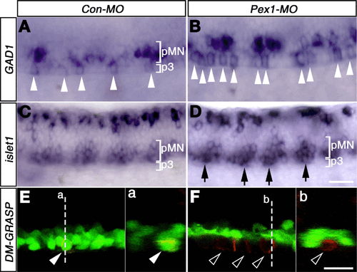
Splice morpholino of PEX1 increases the number of GABAergic KA” neurons. Expression of GAD1 and islet1 by whole-mount in situ hybridization staining in the embryos injected with Con-MO (A, C) and PEX1-MO (B, D) at 25 hpf. Dorsal to the top and anterior to the left. Arrowheads indicate KA” neurons between the 8th and the 10th somite (A, B). Arrows indicate motor neurons in the p3 domain (D). Expression of zn5/DM-GRASP was detected in the red channel of confocal images in Tg[olig2:egfp] embryos injected with Con-MO (E) and PEX1-MO (F) at 27 hpf. Filled arrowheads identify secondary motor neuron (sMN) in the pMN domain of Con-MO-injected Tg[olig2:egfp] embryos (E), and open arrows identify ectopic sMNs in the P3 domain of PEX1-MO-injected embryos (F). Cross-sectional views of a’ and b’, at the position of the dashed lines, are displayed in a and b, respectively. Scale bars: 25 µm. |
|
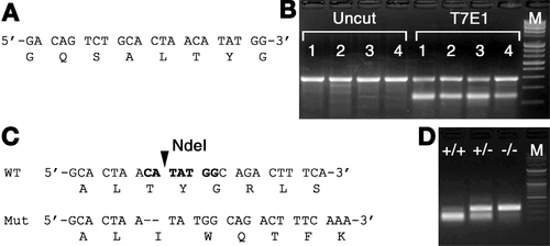
PEX1 mutant is generated by using CRISPR/Cas9 system. (A, B) The efficiency of guide RNA (sgRNA) was analyzed by T7E1 assay. Target sequence of sgRNA for genomic editing was designed based on the exon which has start codon of PEX1 (A). The mixture of synthetic mRNA of Cas9 and sgRNA for PEX1 was microinjected into fertilized eggs of zebrafish. Genomic editing identified in Cas9/sgRNA-injected embryos by T7E1 endonuclease at 24-hpf (B). Uncut represents PCR products without T7E1 digestion. (C, D) Genomic editing was identified in the F2 generation. Sequence analysis confirmed a founder zebrafish which has 2-bp deletion in target sequences (C). The amino acid sequence predicted from the mutated PEX1 sequence and that of wild-type (C). Arrowhead indicate NdeI cleavage site (C). RFLP assay showing the PCR amplicons of PEX1+/+, PEX1+/− and PEX1−/− digested with NdeI restriction enzyme in 24-hpf-old zebrafish embryos (D). M, 100-bp DNA size marker. RFLP, Restriction fragment length polymorphism. |
|
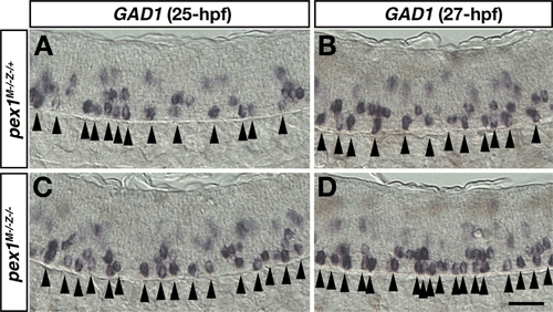
Maternal and zygotic mutations of PEX1 causes an increase in the number of GABAergic KA” neurons in the ventral spinal cord. Whole-mount in situ hybridization staining shows expression of GAD1 in the maternal mutant embryos of PEX1 (A, B) and in the maternal-zygotic mutant embryos of PEX1 (C, D) at 25-hpf (A, C) and 27-hpf (B, D). Arrowheads mark KA” neurons between the 8th- and the 12th-somite (A, C), and between the 8th- and the 11th-somite (B, D). Scale bar; 25 µm. |

