
|
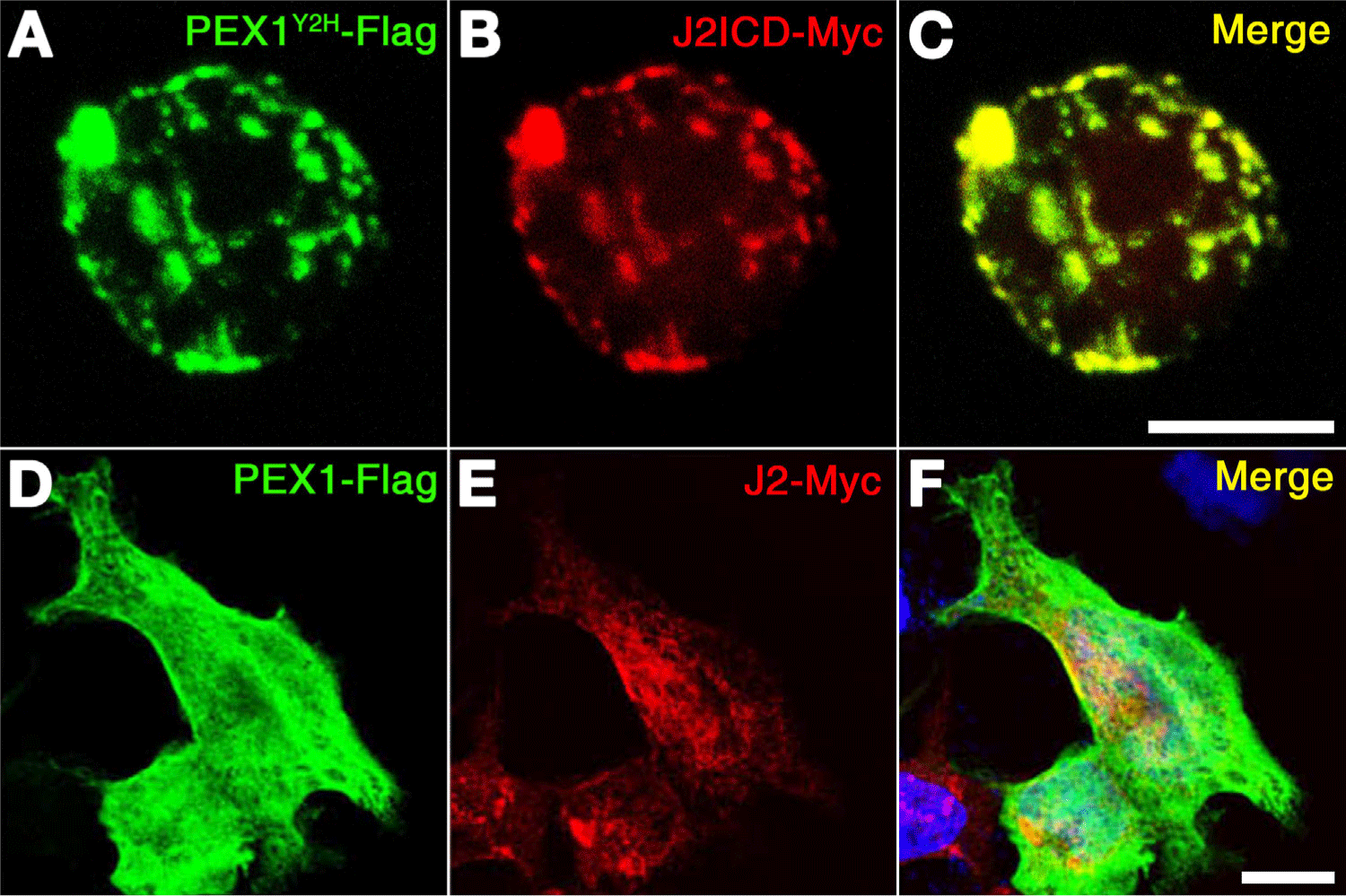
Fig. 2 Zebrafish PEX1 partially co-localized with Jagged2 in the plasma membrane. (A–C) Expression vectors for PEX1Y2H and J2ICD were transfected into P19 cells. Confocal images indicated the Flag-tagged PEX1Y2H in the green channel (A) and the Myc-tagged J2ICD in the red channel (B). The merge image is displayed (C). (D–F) Expression vectors for PEX1 and J2 were transfected into NIH 3T3 cells. Confocal images indicated the Flag-tagged PEX1 in the green channel (D) and the Myc-tagged J2 in the red channel (E). DAPI-stained genomic DNA is shown in the blue channel and the merge image is displayed (F). Scale bars, 10 µm. J2ICD, intracellular domain of zebrafish Jagged2.