The insertional smb mutant presents median fin phenotypes. (A) Whole mount wild-type adult (n=50) and heterozygous smbTg(sox10:Gal4)co3021 (n=50) zebrafish (hereafter smb mutants) were live imaged with transmitted light. In all panels, asterisk denotes the missing dorsal fin; caret indicates reduced size anal fin. (B) Schematic indicating the transgenic sequence and genomic location of the sox10:Gal4 insertion in the smb background. (C) Wild-type and smb heterozygous mutant juveniles (33 dpf) were stained with Alcian Blue (cartilage) and Alizarin Red (bone) and imaged by light microscopy. (D) Enlargements from panel C depicting the radials and rays of the dorsal and anal fins in wild types and mutants. Arrowhead indicates disorganized anal fin radials in mutants. (E) Representative smb heterozygous mutant with partial fin loss was imaged with transmitted light. (F) Arrowheads in enlargements indicate fused radials in reduced dorsal and anal fins. Scale bars: 2 mm (A); 1 mm (C,E).
The chondrogenesis program does not initiate in smb dorsal fins. (A-D) Heterozygous smb mutants were crossed to sox9a:EGFP fish, and offspring were imaged by confocal and transmitted light microscopy during progressive stages of median fin chondrogenesis. (A) In wild-type dorsal fins, the caret indicates initiation of the chondrogenesis program marked by EGFP; white arrowhead indicates stacks of chondrocytes in the developing radials. (B) In smb heterozygous mutants, the asterisk indicates absence of dorsal fin bud mesenchyme and the chondrogenesis program. (C) In wild-type anal fins, the caret indicates initiation of the chondrogenesis program marked by EGFP; white arrowhead indicates stacks of chondrocytes in the developing radials. (D) In smb heterozygous mutant anal fins, the caret indicates initiation of the chondrogenesis program marked by EGFP. White arrowhead indicates stacks of chondrocytes in the developing radials. Fewer individual radials form in mutants. Scale bar: 50 μm.
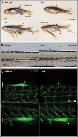
Establishment of dorsal fin bud progenitors is necessary for fin outgrowth. (A) The smb mutant is epistatic to lof. Heterozygous smb adults were crossed to heterozygous lof adults and adult offspring were imaged with transmitted light. The dorsal fin and anal fin are indicated in the wild-type (wt) animal. An asterisk indicates an absent dorsal fin, a caret indicates elongated fins, and guillemets indicate an anal fin that is reduced in the AP axis but elongated in the proximodistal axis in smb;lof double heterozygous mutants (wt=8, smb=4, lof=8, smb;lof=5, expected=6.5 per genotype. Chi-square test; P=0.5641). (B) Nomarski images of fin induction and mesenchyme aggregation in wild type and smb heterozygotes at 5.8 mm SL (n=3 per genotype). Arrowhead indicates fin bud mesenchyme, asterisk marks absent fin bud. (C) Confocal images of live 5.4 mm SL fli1:EGFP wild-type and smb heterozygous mutants (n=10 per genotype). Arrowhead indicates EGFP-expressing mesenchyme cells in the dorsal and anal fin buds. Asterisk indicates absence of fli1:EGFP cells in the dorsal fin bud in smb heterozygous mutants. Scale bars: 3 mm (A); 100 μm (B); 60 μm (C).
The posterior domain of the anal fin is sensitive to the smb insertion. (A) Anterior median fin rays labeled with alx4a:dsRed were live imaged in wild-type and smb heterozygous mutants (n=3 per genotype). (B) Posterior median fin rays labeled with hand2:EGFP were live imaged in wild-type and smb heterozygous mutants (n=3 per genotype). In both panels, asterisk denotes absent dorsal fin, caret indicates reduced anal fin. Scale bars: 150 μm (A); 200 μm (B).
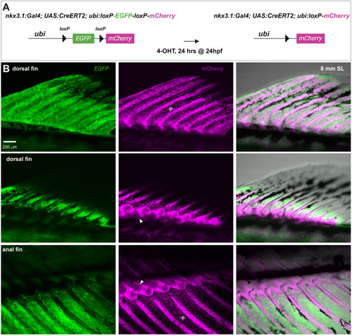
The sclerotome is the source of the dorsal and anal fin skeleton. (A) Schematic indicating transgenic lines and tamoxifen treatment protocol used to lineage trace the sclerotome using nkx3.1:Gal4. (B) Sclerotome-derived, Cre-recombined, mCherry-expressing cells in the dorsal and anal fins were imaged. Arrowheads indicate median fin radial chondrocytes; guillemets indicate median fin ray osteoblasts. n=12/12 individuals with recombination in sclerotome labeled the dorsal and anal fins. Scale bar: 200 μm (B).
smb mutants have impaired sclerotome expansion but early-forming derivatives of the sclerotome are unaffected in the smb mutant. (A) pax9 transcripts were fluorescently labeled with HCR in wild-type and smb heterozygous embryos at 22 and 24 hpf and imaged by confocal microscopy. Gray indicates the pax9+ domain used to quantify volume using Imaris surface rendering. (B) pax9 expression at each condition was quantified by Imaris. For 22 hpf, six wild-type and six smb heterozygous mutant animals were quantified (unpaired two-tailed t-test; P=0.8640; ns, not significant). For 24 hpf, four wild type and four smb heterozygous mutants were quantified. *P=0.0144; unpaired two-tailed t-test. (C) Heterozygous smb fish were crossed to scxa:mCherry fish, and labeled tenocytes in wild-type and smb heterozygous mutant offspring were imaged by confocal microscopy (n=5 per genotype). (D) smb heterozygous fish were crossed to her6:mCherry fish, and fin fold fibroblasts in wild-type and smb heterozygous mutant offspring were imaged by confocal microscopy (n=3 per genotype). (E) Vertebrae were imaged in Alcian Blue and Alizarin Red-stained wild types and stage-matched smb heterozygous mutants. Neural spines (ns), neural arches (na), haemal spines (hs), and haemal arches (ha) are indicated in wild type. Asterisk indicates absent haemal arch and spine; arrowheads mark fused or forked haemal arches or spines. Fisher's exact test; P<0.0001. Scale bars: 50 μm (A, top); 60 μm (A, bottom, C); 30 μm (D); 400 μm (E).
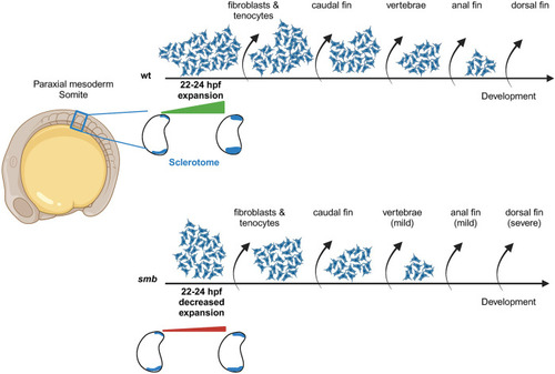
Failed sclerotome expansion and early depletion may underlie fin loss in smb mutants. The sclerotome is specified in the somites at ∼20 hpf, and in wild types expands from 22-24 hpf. Later, this compartment gives rise to multiple tissues in the fish trunk. We propose that as development progresses and tissues are formed, this progenitor pool becomes depleted. In smb, impaired expansion of the sclerotome leads to a smaller initial pool of progenitors, which depletes earlier, leaving no cells to build late-forming structures like the posterior region of the anal fin and the dorsal fin.
Acknowledgments
This image is the copyrighted work of the attributed author or publisher, and
ZFIN has permission only to display this image to its users.
Additional permissions should be obtained from the applicable author or publisher of the image.
Full text @ Development
Your Input Welcome
Thank you for submitting comments. Your input has been emailed to ZFIN curators who may contact you if
additional information is required.
Oops. Something went wrong. Please try again later.