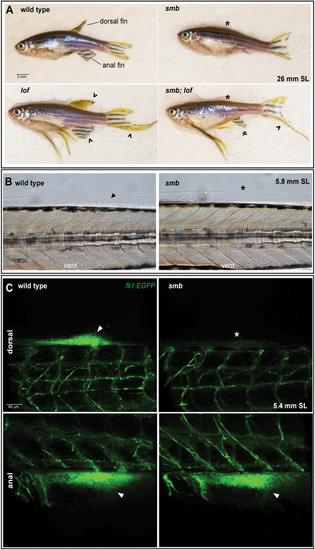
Establishment of dorsal fin bud progenitors is necessary for fin outgrowth. (A) The smb mutant is epistatic to lof. Heterozygous smb adults were crossed to heterozygous lof adults and adult offspring were imaged with transmitted light. The dorsal fin and anal fin are indicated in the wild-type (wt) animal. An asterisk indicates an absent dorsal fin, a caret indicates elongated fins, and guillemets indicate an anal fin that is reduced in the AP axis but elongated in the proximodistal axis in smb;lof double heterozygous mutants (wt=8, smb=4, lof=8, smb;lof=5, expected=6.5 per genotype. Chi-square test; P=0.5641). (B) Nomarski images of fin induction and mesenchyme aggregation in wild type and smb heterozygotes at 5.8 mm SL (n=3 per genotype). Arrowhead indicates fin bud mesenchyme, asterisk marks absent fin bud. (C) Confocal images of live 5.4 mm SL fli1:EGFP wild-type and smb heterozygous mutants (n=10 per genotype). Arrowhead indicates EGFP-expressing mesenchyme cells in the dorsal and anal fin buds. Asterisk indicates absence of fli1:EGFP cells in the dorsal fin bud in smb heterozygous mutants. Scale bars: 3 mm (A); 100 μm (B); 60 μm (C).
|
