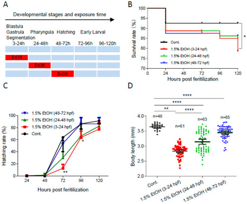
The temporal window of ethanol sensitivity in zebrafish embryos. (A) Zebrafish embryos were exposed to 1.5% ethanol during either 3–24 hpf, 24–48 hpf, or 48–72 hpf, then rinsed and incubated in egg water. The survival rate, hatching rate, and body length of exposed groups and control were indicated in (B–D), respectively. One-way ANOVA was used to compare the ethanol exposure groups to the control with * p < 0.05, ** p < 0.01, and **** p < 0.0001.
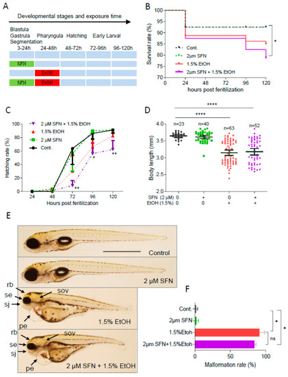
Pretreatment with SFN showed no protective effect against ethanol-induced teratogenicity. (A) Zebrafish embryos were treated with 2 µM SFN from 3 to 24 hpf, then exposed to 1.5% ethanol from 24 to 48 hpf, and thereafter incubated in fresh egg water. (B) Survival rate. (C) Hatching rate. (D) Body length. (E) Representative larvae of control without treatment (first row), 3–24 hpf treatment with 2 µM SFN (second row from top), 24–48 exposure with 1.5% ethanol (third row), and 3–24 hpf pretreatment with 2 µM SFN and then 24–48 exposure with 1.5% ethanol (bottom row). (F) Histogram (next to the larvae images) indicates the percentage of larvae with typical FASD malformations. Arrows indicate the phenotypes of FASD. se, small eyes; rb, reduced brain; sov, small otic vesicle; sj, small jaw; and pe, pericardial edema. One-way ANOVA was used to analyze the effects of SFN alone and t-test for pairwise comparison with ns, not significant, * p < 0.05, ** p < 0.01, and **** p < 0.0001. The scale bar represents 1 mm.
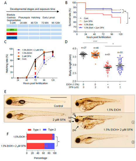
Co-treatment with SFN protected against ethanol-induced teratogenicity. (A) Zebrafish embryos were co-treated with 2 µM SFN, 1.5% ethanol during 3–24 hpf. (B) Survival rate. (C) Hatching rate. (D) Body length. (E) Representative larvae of control without treatment, 3–24 hpf treatment with 2 SFN alone, 3–24 hpf treatment with 1.5% ethanol column, and co-treatment with SFN and ethanol. The larvae in co-treatment groups were classified into two types: type 1 larvae are typically FASD-like, whereas type 2 larvae are more similar to SFN-supplemented control. (F) Percentage of type 1 and 2 larvae in the co-treatment groups. Arrows indicate the phenotypes of fetal alcohol spectrum disorder (FASD). se, small eyes; rb, reduced brain; sov, small otic vesicle; sj, small jaw; and pe, pericardial edema. One-way ANOVA was used to analyze the effects of SFN alone and t-test for pairwise comparison with ns, not significant, * p < 0.05, ** p < 0.01, and **** p < 0.0001. The scale bar represents 1 mm.
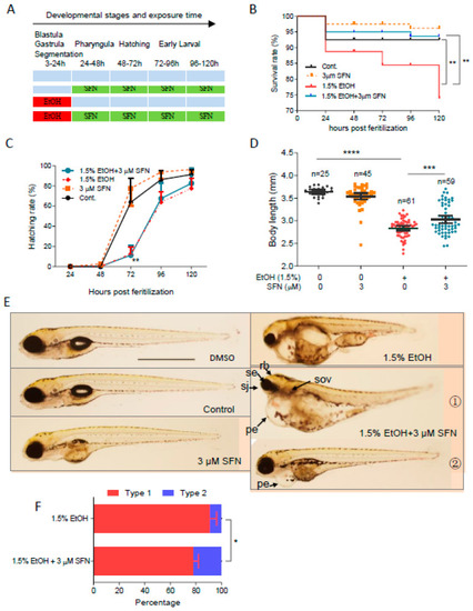
Post-treatment with SFN also rescued FASD in zebrafish embryos. (A) Zebrafish embryos were first exposed to 1.5% ethanol during 3–24 hpf and then treated with 3 µM SFN for 96 h with renewal every 24 h. Treatment with 3 µM SFN alone with renewal every 24 h and treatment with 1.5% ethanol alone during 3–24 hpf were also performed. (B) Survival rate. (C) Hatching rate. (D) Body length. (E) Representative larvae of DMSO control and freshwater control without treatment, 24–120 hpf treatment with 3 µM SFN alone, 3–24 hpf treatment with 1.5% ethanol, and post-treatment with SFN and ethanol. The larvae in post-treatment groups were claasified into two types: type 1 larvae are typically FASD-like, whereas type 2 larvae are more like SFN-supplemented control. (F) Percentage of type 1 and 2 larvae in the post-treatment groups. Arrows indicate the phenotypes of fetal alcohol spectrum disorder (FASD). se, small eyes; rb, reduced brain; sov, small otic vesicle; sj, small jaw; and pe, pericardial edema. One-way ANOVA was used to analyze the effects of SFN alone and t-test for pairwise comparison with * p < 0.05, ** p < 0.01, *** p < 0.001, and **** p < 0.0001. The scale bar represents 1 mm.
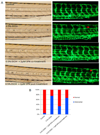
Co-treatment and post-treatment of SFN attenuate ethanol-induced dysangiogenesis in zebrafish embryos. (A) Zebrafish embryos (Tg: fli1-eGFP) were exposed to 0.5% ethanol during 3–24 hpf with or without 2 μM SFN during the same development phase. A group of embryos were also post-treated with 2 μM SFN after being exposed to 0.5% ethanol during 3–24 hpf. Representative images show the vascular structure in the trunk area of the embryos. Arrows indicate the malformation of the intersegmental vessels. (B) The bar graph shows the percentage of embryos with or without vessel malformation.
Acknowledgments
This image is the copyrighted work of the attributed author or publisher, and
ZFIN has permission only to display this image to its users.
Additional permissions should be obtained from the applicable author or publisher of the image.
Full text @ Int. J. Mol. Sci.
Your Input Welcome
Thank you for submitting comments. Your input has been emailed to ZFIN curators who may contact you if
additional information is required.
Oops. Something went wrong. Please try again later.