FIGURE SUMMARY
Title

A dTDP-L-Rhamnose 4-epimerase required for glycopeptidolipid biosynthesis in Mycobacterium abscessus

Authors
Aguilera-Correa, J.J., Wei, F., Leclercq, L.D., Tasrini, Y., Mullapudi, E., Daher, W., Nakajima, K., Canaan, S., Herrmann, J.L., Wilmanns, M., Guérardel, Y., Wen, L., Kremer, L.
Source
Full text @ J. Biol. Chem.

MAB_4111c belongs to the GPL biosynthetic cluster and is a predicted epimerase.A, structure of diglycosylated (GPL-2a) and triglycosylated (GPL-3) glycopeptidolipids. The green, blue, and yellow shadings represent the lipid, peptide, and glycosydic moieties of the GPL. Di-O-acetyl 6-d-Tal is drawn in red. B, Mycobacterium abscessus gpl locus encoding enzymes involved in the synthesis, modification, and transport of GPL. MAB_4111c is indicated in bold. C, alignment of the protein sequence of dTDP-L-Rha 4-epimerase from the Burkholderia thailandensis (ATCC 700388) (wbiB_BURTA) with that of MAB_4111c from M. abscessus. The orange and blue shadings represent the nonconserved and conserved and amino acids, respectively. The red, green, and blue asterisks indicate residues involved in the putative NADH-binding site, the substrate-binding site, and the catalytic residue reported in B. thailandensis, respectively.

Enzymatic activity of MAB_4111c.A, reaction catalyzed by MAB_4111c. B, upper panel: HPLC profile of the standards and products formed during the enzymatic reaction in the presence of MAB_4111c or MAB_4111c_Y181A with dTDP-Rha and NADP+. The incubation was performed for 60 min. The product of the reaction is labeled with a red box. Lower panel: HPLC profile of standard dTDP-Glc, standard dTDP-6-d-Tal, and dTDP-Glc, and NADP+ incubated with MAB_4111c for 60 min. C, upper panel: LC/MS profile of the enzymatic reaction mixture in MRM mode using the specific precursor ion [M-H] at m/z 547.1. Lower panel: MS2 fragmentation spectra of standard dTDP-6-d-Tal and the enzymatic reaction product (∗). D, 1H NMR spectra of dTDP-L-Rha and NADP+; MAB_4111c with dTDP-L-Rha and NADP+ after 120 min; MAB_4111c_Y181A with dTDP-L-Rha and NADP+ after 120 min; dTDP-6-d-Tal. The characteristic peaks of dTDP-6-d-Tal are indicated with red arrows. E, kinetic analysis of the reaction catalyzed by MAB_4111c in the presence of dTDP-L-Rha and NADP+.

Structural prediction of Tle.A, cartoon representation of the Alphafold 3 predicted dimeric structure of Tle. The structure is colored according to the plDTT score range (blue = high confidence, cyan = confidence, yellow = low confidence, orange = very low confidence). B, left panel: cartoon representation of the predicted Tle homodimer, with each chain colored in green and cyan. NAD is depicted as a stick model. Right panel: alignment of the monomeric structure of Tle with the WbpP crystal structure (Tle in cyan and WbpP in magenta).

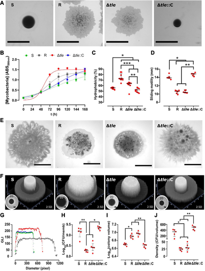
Deletion of tle in M. abscessus alters colony morphology, hydrophobicity, sliding motility, and biofilm-colony development.A, knock-out of tle is associated with a switch from an S to an R-like morphotype. The scale bar represents 1 mm. B, the deletion of tle is associated with a higher growth rate. The experiment was performed using two biological replicates with six technical replicates per biological replicate. Deletion of tle modifies hydrophobicity (C) and the sliding motility (D and E). The hydrophobicity experiment was performed using three biological replicates with three technical replicates per biological replicate. The sliding motility experiment was performed using three biological replicates with two technical replicates per biological replicate. The black scale bar indicates 5 mm. F, representative 2- and 2.5-dimensional pictures of colony-biofilms on biofilm-supporting membranes after 5 days of incubation. The absence of tle alters the biofilm-colony growth (F) (the scale bar represents 2 mm) and the biofilm-colony profile (G) (green, dark grey, red, and blue dots represent the profiles of S, R, Δtle and Δtle::C, respectively). The CFU per membrane are shown in (H), the colony volume in (I), and the colony density in (J). This experiment was performed using five biological replicates. The error bar denotes the interquartile range. ∗: p < 0.05, ∗∗: p < 0.01, and ∗∗∗: p < 0.001. CFU, colony-forming units.

Structural analysis of the GPL content in Δtle.A, TLC analysis representative from three independent experiments of native apolar (left) and polar (right) lipid extracts. Lipids were separated using chloroform/methanol/water (90:10:1, v/v/v) followed by spraying with orcinol and charring. GPL-1b and GPL-2b previously described (35) from Δtle display a lower Rf than WT GPL-2a and GPL-3, which are restored in Δtle::C. TDM: trehalose dimycolate; DAT: Di-O-acyl trehalose. B, TLC analysis of native (NaOH−) and saponified (NaOH+) polar fractions. Lipids were separated using chloroform/methanol/water (90:10:1, v/v/v) followed by spraying with orcinol and charring. The Rf of GPL-2a and GPL-3 are identical in S and Δtle::C but not in Δtle. C, MALDI-TOF positive MS spectra of native polar lipid fraction (black, NaOH−) from the S and Δtle::C strains show ions for GPL-2a at m/z 1257.9/1285.9 and for GPL-3 at m/z 1403.9/1432.0. MS spectra from saponified polar lipid fraction (red, NaOH+) show a 84 u.m.a decrease, to 1173.4/1201.4 and 1319.3/1347.4, corresponding to deacetylated dGPL-2a and dGPL-3, as previously described (34). Conversely, ions for GPL-1b at m/z 1013.8/1041.8 and from GPL-2b at 1187.9 were intense in native and saponified polar extracts from Δtle. D, relative proportion of each monosaccharides was determined in the GPL-enriched saponified polar lipid fraction. 6-d-Tal could only be detected in S and Δtle::C.

Distribution of tle orthologs in nontuberculous mycobacteria and correlation with the presence of 6-d-Tal.A, phylogenetic tree reconstructed using already available tle sequences. Nonmycobacterial species and mycobacterial species are represented in pink and blue areas, respectively. Green, yellow, and red dots label slowly-growing, intermediately-growing, and rapid-growing mycobacteria, respectively. B, TLC analysis of the polar lipid fraction of a selected panel of NTM clinical strains, representative from two independent experiments. Lipids were separated using chloroform/methanol/water (90:10:1, v/v/v) followed by spraying with orcinol and charring. +: presence of GPL-like components, confirmed by their detection after saponification (Fig. S8). −: absence of GPL-like components. C, Deoxyhexose composition analysis of polar lipid fractions by GC/FID and GC/MS. Relative area of identified monosaccharides (n = 4) show Rha residues in green and 6-d-Tal in blue. Identification of 6-d-Tal and Rha was confirmed by the retention time and fragmentation pattern of standards while O-methylated Rha was determined previously (33, 35). Identification of 2- and 3-O-methyl 6-d-Tal was deduced from the retention time and fragmentation pattern. Relative areas of Rha and 6-d-Tal and their methylated forms are expressed as ratios of total monosaccharides.

Increased virulence and pathogenesis of Δtle in a zebrafish embryo model of infection.A, schematic overview of in vivo experiments. B, survival of infected embryos with the R strain (275 ± 27 CFU), the S strain (280 ± 36 CFU), Δtle (264 ± 33 CFU), and Δtle::C (281 ± 47 CFU). The graph shows the result of four pooled independent experiments. Embryos were marked dead in the absence of a heartbeat. Statistical analysis was performed using Log-rank (Mantel-Cox) test. ∗∗∗∗: p < 0.0001; ∗∗: p < 0.01, ns: non-significant. C, bacterial burden from infected embryos at 2, 3, and 5 dpi. The graph shows the result of three pooled independent experiments. n = 5 embryos per time-point per condition. CFU counts were Log10 transformed. ∗: p < 0.05, ∗∗: p < 0.01, ∗∗∗: p < 0.001, and ∗∗∗∗: p < 0.0001. D, imaging of infected embryos at 3 dpi. One zoomed representative image of the head is shown per condition. Scale bars represent 0.2 mm. CFU, colony-forming units.

The biosynthetic interconnections between the arabinogalactan and glycopeptidolipid pathways in M. abscessus. dTDP-L-Rha is generated by the successive action of the Rml pathway enzymes, consisting of RmlA (MAB_4113), RmlB (MAB_3779), RmlC (MAB_3780), and RmlD (MAB_3613c). This dTDP-L-Rha is used by WbbL (MAB_3612c) to produce the Rha-N-acetyl-glucosaminyl-phosphate disaccharide unit that cross-links arabinogalactan to peptidoglycan. In addition, dTDP-L-Rha is also used by Gtf2 (MAB_4104) and Gtf3 (MAB_4112c) for GPL rhamnosylation (blue arrows). dTDP-6-d-Tal, produced from dTDP-L-Rha by the dTDP-Rha-4-epimerase (MAB_4111c/Tle) serves as the substrate for the talosyltransferase Gtf1 (green arrows), which adds one 6-d-Tal unit to mono- or di-rhamnosylated GPL precursors. The glycosylated GPL (GPL-2a and GPL-3) are then transported across the inner membrane by the MmpL4 complex (MAB_4116c, MAB_4115c, MAB_4117c) and inserted into the outer leaflet of the mycomembrane in the M. abscessus S variant.

Acknowledgments
This image is the copyrighted work of the attributed author or publisher, and ZFIN has permission only to display this image to its users. Additional permissions should be obtained from the applicable author or publisher of the image. Full text @ J. Biol. Chem.