
- Title
-
A novel NIR fluorescent probe for visualizing hydrogen sulfide in Alzheimer's disease
- Authors
- Hong, S., Gan, Y., Liu, D., Yu, T., Zhou, H., Li, H., Liu, F., Yin, P.
- Source
- Full text @ Analyst
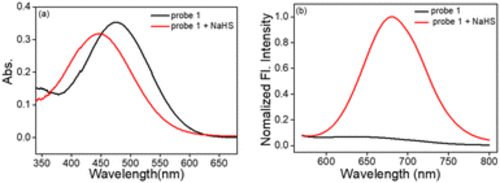
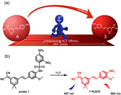
(a) Absorption spectra of probe 1 (10 μM) upon addition of 10 equiv. of NaHS in DMSO–PBS (10 mM, pH = 7.4, v/v, 1/9) at 37 °C for 60 min. (b) Normalized fluorescence spectra of probe 1 (10 μM) upon addition of 10 equiv. of NaHS in DMSO–PBS (10 mM, pH = 7.4, v/v, 1/9) at 37 °C for 60 min. λex = 457 nm, λem = 680 nm. Slit (nm): 5.0/10.0. |
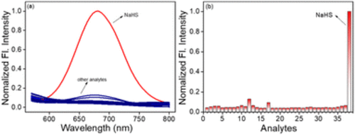
(a) The fluorescence spectra of probe 1 (10 μM) in the presence of Cys (200 μM), GSH (200 μM), and other analytes (100 μM) in DMSO–PBS (pH 7.4, 10 mM, v/v, 1/9) at 37 °C. Each spectrum was recorded after 60 min. λex = 457 nm, slit (nm): 5/10.0. Analytes: (1) probe 1, (2) H2O2, (3) ONOO−, (4) NO, (5) O2˙−, (6) 1O2, (7) tBuOO−, (8) NaHSO3, (9) NAC, (10) Hcy, (11) GSH, (12) Cys, (13) Na2S2, (14) Sec, (15) Na+, (16) Ag+, (17) K+, (18) Co2+, (19) Zn2+, (20) Fe2+, (21) Ca2+, (22) Ba2+, (23) Val, (24) His, (25) Pro, (26) Gly, (27) Lys, (28) Glu, (29) Ser, (30) Thr, (31) Asp, (32) Arg, (33) Leu, (34) Trp, (35) HPO42−, (36) H2PO4−, (37) HCO3−, (38) NaHS. (b) The corresponding fluorescence intensity change at 680 nm of probe (10 μM) to other analytes (100 μM) in DMSO–PBS (10 mM, pH = 7.4, v/v, 1/9). |
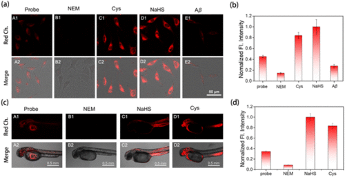
(a) Confocal fluorescence imaging of endogenous and exogenous H2S in SH-SY5Y cells. (A1 and A2) Cells were incubated with probe 1 (5 μM) for 30 min. (B1 and B2) The cells were pretreated with NEM (200 μM) for 30 min, and then incubated with probe 1 (5 μM, 30 min). (C1 and C2) The cells were pretreated with Cys (200 μM) for 30 min, and then incubated with probe 1 (5 μM, 30 min). (D1 and D2) Cells were pretreated with NEM (0.2 mM) for 30 min, subsequently incubated with NaHS (100 μM, 30 min), and finally incubated with probe 1 (5 μM) for 30 min. (E1 and E2) Cells were pretreated with Aβ42 aggregates (5 μM) for 60 min and then incubated with probe 1 (5 μM) for 30 min. (b) Relative pixel intensity in panel (a) (λex = 458 nm, λem = 600–750 nm for the red channel). Scale bar: 50 μm. (c) Confocal fluorescence imaging of endogenous and exogenous H2S in zebrafish. (A1 and A2) Zebrafish were incubated with probe 1 (10 μM) for 60 min. (B1 and B2) The cells were pretreated with NEM (200 μM) for 30 min, and then incubated with probe 1 (10 μM, 60 min). (C1 and C2) Cells were pretreated with NEM (200 μM) for 30 min, subsequently incubated with NaHS (100 μM, 30 min), and finally incubated with probe 1 (10 μM) for 60 min. (D1 and D2) The cells were pretreated with Cys (100 μM) for 30 min, and then incubated with probe 1 (10 μM, 60 min). (d) Relative pixel intensity in panel (c) (λex = 458 nm, λem = 600–750 nm for the red channel). Scale bar: 0.5 mm. Data are mean ± SEM, n = 3. |
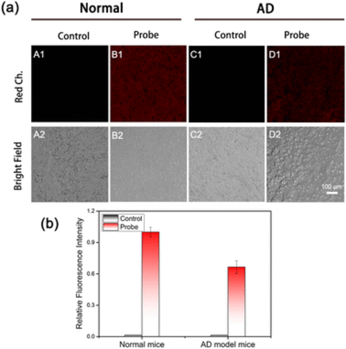
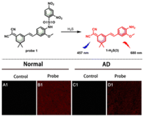
(a) Confocal fluorescence images of endogenous H2S in hippocampus tissues slices from normal and AD model mice. (A1, A2 and C1, C2) The fresh hippocampus tissues from normal and AD mice pretreated with PBS (30 min). (B1, B2 and D1, D2) Hippocampus tissues from normal and AD mice pretreated with probe 1 (10 μM, 60 min). (b) Relative pixel intensity in panel (a) (λex = 458 nm, λem = 600–750 nm for the red channel). Scale bar: 100 μm. Data are mean ± SEM, n = 3. |
|
|