
Fig. 2
- ID
- ZDB-FIG-240822-25
- Publication
- Hong et al., 2024 - A novel NIR fluorescent probe for visualizing hydrogen sulfide in Alzheimer's disease
- Other Figures
- All Figure Page
- Back to All Figure Page
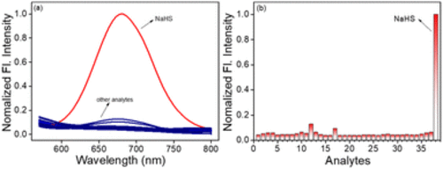
(a) The fluorescence spectra of probe 1 (10 μM) in the presence of Cys (200 μM), GSH (200 μM), and other analytes (100 μM) in DMSO–PBS (pH 7.4, 10 mM, v/v, 1/9) at 37 °C. Each spectrum was recorded after 60 min. λex = 457 nm, slit (nm): 5/10.0. Analytes: (1) probe 1, (2) H2O2, (3) ONOO−, (4) NO, (5) O2˙−, (6) 1O2, (7) tBuOO−, (8) NaHSO3, (9) NAC, (10) Hcy, (11) GSH, (12) Cys, (13) Na2S2, (14) Sec, (15) Na+, (16) Ag+, (17) K+, (18) Co2+, (19) Zn2+, (20) Fe2+, (21) Ca2+, (22) Ba2+, (23) Val, (24) His, (25) Pro, (26) Gly, (27) Lys, (28) Glu, (29) Ser, (30) Thr, (31) Asp, (32) Arg, (33) Leu, (34) Trp, (35) HPO42−, (36) H2PO4−, (37) HCO3−, (38) NaHS. (b) The corresponding fluorescence intensity change at 680 nm of probe (10 μM) to other analytes (100 μM) in DMSO–PBS (10 mM, pH = 7.4, v/v, 1/9). |