Image
Figure Caption
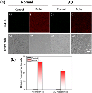
Fig. 4 (a) Confocal fluorescence images of endogenous H2S in hippocampus tissues slices from normal and AD model mice. (A1, A2 and C1, C2) The fresh hippocampus tissues from normal and AD mice pretreated with PBS (30 min). (B1, B2 and D1, D2) Hippocampus tissues from normal and AD mice pretreated with probe 1 (10 μM, 60 min). (b) Relative pixel intensity in panel (a) (λex = 458 nm, λem = 600–750 nm for the red channel). Scale bar: 100 μm. Data are mean ± SEM, n = 3.
Acknowledgments
This image is the copyrighted work of the attributed author or publisher, and
ZFIN has permission only to display this image to its users.
Additional permissions should be obtained from the applicable author or publisher of the image.
Full text @ Analyst

