
- Title
-
Oxysterol-binding protein like 7 deficiency leads to ER stress mediated apoptosis in podocytes and proteinuria
- Authors
- Duara, J., Torres, M.F., Gurumani, M., Molina David, J., Njeim, R., Kim, J.J., Mitrofanova, A., Ge, M., Sloan, A.J., Müller-Deile, J., Schiffer, M., Merscher, S., Fornoni, A.
- Source
- Full text @ Am. J. Physiol. Renal Physiol.
Decreased OSBPL7 expression in mouse models of experimental CKD is associated with podocyte injury. A and B: representative Western blot images (A) and quantification showing OSBPL7 levels in the kidney cortex of Col4a3−/− and db/db mice compared with Col4a3+/+ (n = 3, *P < 0.05) and db/+ (n = 4, **P < 0.001) littermates (B). C: immunofluorescence staining showcasing OSBPL7 (green) and WT1 (red) in glomeruli, with the merged image highlighting their colocalization, indicating OSBPL7's association with podocytes. D: quantification of OSBPL7 and WT1 colocalization, presented as the ratio of OSBPL7/WT1 double-positive signals to total WT1-positive cells, showing the significant decrease in OSBPL7 in the glomeruli of CKD models compared with controls (n = 10, ****P < 0.0001). E: stable OSBPL7-deficient (SiOSBPL7) podocytes were generated using siRNA. F: apoptosis levels increased in OSBPL7-deficient podocytes and returned to control levels with transfection of full-length OSBPL7 (OSBPL7FL) but not with transfection of OSBPL7 plasmid containing a deletion of the FFAT domain (OSBPL7FFAT−). n = 3 technical replicates. *P < 0.05 and **P < 0.01. G: immunofluorescent images of OSBPL7 (red), ER stain (green), and DAPI (blue) in siOSBPL7 cells with and without OSBPL7FL or OSBPL7FFAT− expressing plasmid. CKD, chronic kidney disease; OSBPL7, oxysterol-binding protein-like 7. |
OSBPL7 deficiency leads to ER stress and decreased autophagic flux. A and B: representative Western blot images (A) of PERK, IRE1α, PDI, phosphorylated and total SAPK/JNK, BiP, and GAPDH with quantification from three independent experiments (B). C and D: representative Western blot images (C) of LC3I, LC3II, and GATE16 levels with GAPDH loading control with quantification from three independent experiments (D). n = 3. *P < 0.05, **P < 0.01, and ***P < 0.005. BiP, binding immunoglobulin protein; ER, endoplasmic reticulum; IRE1α, inositol-requiring enzyme 1α; OSBPL7, oxysterol-binding protein-like 7; PERK, protein kinase RNA-like ER kinase; SAPK/JNK, stress-activated protein kinase/c-Jun N-terminal kinase. |
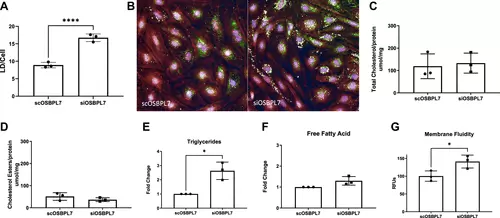
OSBPL7 deficiency in podocytes alters lipid homeostasis. A: LD quantification per cell in siOSBPL7 podocytes versus control. B: representative images of LD in scOSBPL7 and siOSBPL7 cells stained for LD (green), cytoskeleton (red), and nucleus (blue). Total cholesterol (C) and cholesterol ester levels (D) were not changed between siOSBPL7 and scOSBPL7 podocytes. Triglycerides were increased in siOSBPL7 podocytes (E), while free fatty acids (F) were not changed as indicated by the fold change from siOSBPL7 compared with scOSBPL7 levels. G: membrane fluidity was increased in siOSBPL7 podocytes compared with scOSBPL7. n = 3. *P < 0.05 and ****P < 0.001. LD, lipid droplet; OSBPL7, oxysterol-binding protein-like 7. |
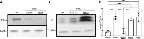
ER stress and not decreased autophagy or increased LD accumulation is responsible for apoptosis in podocytes. A: Western blot analysis illustrating the effect of KIRA6 on IRE1α expression in siOSBPL7 podocytes, with a marked reduction in IRE1α levels observed. The underlined and bold concentration indicates the specific concentration used in our experiments. B: Western blot showing the impact of STF62247 on LC3 levels in siOSBPL7 podocytes. The increase in LC3 levels upon STF62247 treatment confirmed the promotion of autophagy. The underlined and bold concentration indicates the specific concentration used in our experiments. C: apoptosis assessment in siOSBPL7 podocytes posttreatment with KIRA6, STF62247, and hydroxypropyl β-cyclodextrin (CD). Apoptosis, heightened in untreated cells, was normalized with KIRA6 treatment. STF62247 and CD did not significantly mitigate apoptosis, highlighting ER stress as the central mechanism in apoptosis induction, rather than autophagy or LD content alterations. n = 3. ***P < 0.005. ER, endoplasmic reticulum; IRE1α, inositol-requiring enzyme 1α; LC-3, light chain-3; LD, lipid droplet; OSBPL7, oxysterol-binding protein-like 7. |
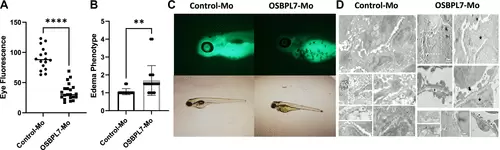
Proteinuria and glomerular damage in zebrafish with Osbpl7 knockdown. Osbpl7 knockdown by osbpl7-Mo in l-fabp:DBP-eGFP ZF led to decreased eye fluorescence (A) and increased edema (B). C: representative images of individual zebrafish. D: transmission electron microscopy (TEM) images of the zebrafish glomerulus at 5 DPI with either Control-Mo and osbpl7-Mo. Substantial glomerular damage was conspicuous in the osbpl7-Mo-injected larvae. The detailed TEM analysis revealed the loss of endothelial fenestrations (white arrows) and widening and rupture of the glomerular basement membrane (GBM) (star). The distinct alterations in podocyte structures are shown, including podocyte effacement and protrusions of foot processes into the GBM (black arrows), a common hallmark of proteinuric states in fish. These observations point to clear podocyte and glomerular damage, in alignment with the proteinuria documented in the fish. 4 DPI. n = 16. **P < 0.01 and ****P < 0.001. OSBPL7, oxysterol-binding protein-like 7. |

ZFIN is incorporating published figure images and captions as part of an ongoing project. Figures from some publications have not yet been curated, or are not available for display because of copyright restrictions. |