
Fig. 4
- ID
- ZDB-IMAGE-240828-4
- Publication
- Duara et al., 2024 - Oxysterol-binding protein like 7 deficiency leads to ER stress mediated apoptosis in podocytes and proteinuria
- All Figures
- Figures for Duara et al., 2024
|
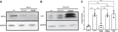
Fig. 4 ER stress and not decreased autophagy or increased LD accumulation is responsible for apoptosis in podocytes. A: Western blot analysis illustrating the effect of KIRA6 on IRE1α expression in siOSBPL7 podocytes, with a marked reduction in IRE1α levels observed. The underlined and bold concentration indicates the specific concentration used in our experiments. B: Western blot showing the impact of STF62247 on LC3 levels in siOSBPL7 podocytes. The increase in LC3 levels upon STF62247 treatment confirmed the promotion of autophagy. The underlined and bold concentration indicates the specific concentration used in our experiments. C: apoptosis assessment in siOSBPL7 podocytes posttreatment with KIRA6, STF62247, and hydroxypropyl β-cyclodextrin (CD). Apoptosis, heightened in untreated cells, was normalized with KIRA6 treatment. STF62247 and CD did not significantly mitigate apoptosis, highlighting ER stress as the central mechanism in apoptosis induction, rather than autophagy or LD content alterations. n = 3. ***P < 0.005. ER, endoplasmic reticulum; IRE1α, inositol-requiring enzyme 1α; LC-3, light chain-3; LD, lipid droplet; OSBPL7, oxysterol-binding protein-like 7.