FIGURE SUMMARY
Title

The splicing factor Prpf31 is required for hematopoietic stem and progenitor cell expansion during zebrafish embryogenesis

Authors
Lv, Y., Li, J., Yu, S., Zhang, Y., Hu, H., Sun, K., Jia, D., Han, Y., Tu, J., Huang, Y., Liu, X., Zhang, X., Gao, P., Chen, X., Shaw Williams, M.T., Tang, Z., Shu, X., Liu, M., Ren, X.
Source
Full text @ J. Biol. Chem.

Expression patterns of prpf31 during zebrafish embryonic development in siblings and prpf31−/−zebrafish.AL and A’L’, prpf31 expression in siblings and prpf31−/− zebrafish was examined by WISH at different developmental stages. EL and E’L’: lateral views, anterior to the left and dorsal upward. G and G’: magnified views of the ICM in (E and E’). H and H’: magnified views of the PBI in (F and F’). K, L, K’, and L’: magnified views of the CHT in (I, J, I’, and J’). The number of embryos with similar gene expression patterns among all embryos examined was shown at the bottom right of each panel. The scale bars represent 250 μm. CHT, caudal hematopoietic tissue; HSPC, hematopoietic stem and progenitor cell; ICM, intermediate cell mass; PBI, posterior blood island; PRPF, pre-mRNA processing factor; WISH, whole-mount in situ hybridization.

Definitive hematopoiesis is impaired in prpf31−/−zebrafish.A, bright field observation showed morphological abnormalities in prpf31−/− zebrafish at 3 dpf. WISH manifested the expression of HSPC markers runx1 and cmyb were noticeably reduced and rescue of cmyb expression after prpf31 mRNA injection. Lateral views. B, expression of the myelocyte markers lyz, l-plastin, and mpx by WISH, and staining signal of Sudan black B labeled neutrophils were significantly decreased in the CHT of prpf31−/− zebrafish at 3 dpf. Lateral views. C, expression of the erythrocyte markers hbae1.1, hbae3, and hbbe1 were almost undetectable in the CHT of prpf31−/− zebrafish at 4 dpf, although there was only slightly decrease compared with siblings at 3 dpf by WISH. Lateral views. D, expression of the early T cell marker rag1 was almost completely absent in the thymus of prpf31−/− zebrafish at 3 dpf by WISH. Lateral and dorsal views. Black arrows denote the CHT region. Red arrows denote the thymus region. The number of embryos with similar gene expression patterns among all embryos examined was shown at the bottom right of each panel. The scale bars represent 250 μm. E, quantification of mRNA signals of prpf31−/− zebrafish and siblings detected by WISH in (AD). The total number of embryos examined was indicated below each column. Mean ± SD; unpaired two-tailed t test; ∗p < 0.05, ∗∗p < 0.01, ∗∗∗p < 0.001; ns, not significant. CHT, caudal hematopoietic tissue; dpf, days postfertilization; hpf, hours postfertilization; HSPC, hematopoietic stem and progenitor cell; PRPF, pre-mRNA processing factor; WISH, whole-mount in situ hybridization.

HSPC expansion and maintenance in the CHT are compromised in prpf31−/−zebrafish.AI and A’–I’, time-course analysis of the expression of HSPC markers runx1 and cmyb in prpf31−/− and sibling embryos from 28 hpf to 48 hpf by WISH. The reduced expression of cmyb in prpf31−/− zebrafish was rescued by prpf31 mRNA injection at 48 hpf. Lateral views. Red arrows denote the AGM region. Black arrows denote the CHT region. The number of embryos with similar gene expression patterns among all embryos examined was shown at the bottom right of each panel. The scale bars represent 250 μm. J, quantification of mRNA signals of prpf31−/− zebrafish and siblings detected by WISH in (AI and A’I’). The total number of embryos examined was indicated below each column. Mean ± SD; unpaired two-tailed t test; ∗p < 0.05, ∗∗p < 0.01, and ∗∗∗p < 0.001; ns, not significant. K, in vivo observation of HSPCs using transgenic line Tg (cmyb: EGFP) of prpf31−/− zebrafish and siblings, in the AGM and CHT at 36 hpf, 48 hpf, and 3 dpf. The total number of embryos examined was indicated at the bottom right of each panel. The scale bars represent 50 μm. L, quantification of the number of HSPCs in the AGM and CHT of prpf31−/− zebrafish and siblings observed in (K). The total number of embryos examined was indicated below each column. Mean ± SD; unpaired two-tailed t test; ∗p < 0.05, ∗∗∗p < 0.001; ns, not significant. AGM, aorta-gonad-mesonephros; CHT, caudal hematopoietic tissue; dpf, days postfertilization; hpf, hours postfertilization; HSPC, hematopoietic stem and progenitor cell; PRPF, pre-mRNA processing factor; WISH, whole-mount in situ hybridization. EGFP, enhanced green fluorescent protein.

HSPC proliferation in the CHT of prpf31−/−zebrafish is arrested in M phase.A, double staining of cmyb:EGFP and EdU showed no obvious difference of EdU+ HSPCs in the AGM and CHT at 36 and 48 hpf. The total number of embryos examined were indicated at the top right of each panel. The scale bars represent 50 μm. B, quantification of the percentage and number of EdU+ HSPCs was detected in (A). The total number of embryos examined was indicated below each column. Mean ± SD; unpaired two-tailed t test; ∗p < 0.05, ∗∗p < 0.01; ns, not significant. C, double immunostaining of cmyb:EGFP and pH3 (Ser10) showed a significant increase of pH3+ HSPCs in the CHT at 48 hpf, but not in the AGM and CHT at 36 hpf. The total number of embryos examined were indicated at the top right of each panel. The scale bars represent 50 μm. D, quantification of the percentage and number of pH3+ HSPCs was detected in (C). The total number of embryos examined was indicated below each column. Mean ± SD; unpaired two-tailed t test; ∗p < 0.05, ∗∗∗p < 0.001; ns, not significant. AGM, aorta-gonad-mesonephros; CHT, caudal hematopoietic tissue; EdU, 5-ethynyl-29-deoxyuridine; hpf, hours postfertilization; HSPC, hematopoietic stem and progenitor cell; pH3, phospho-histone 3. EGFP, enhanced green fluorescent protein.

prpf31−/−zebrafish exhibit severe aberrant alternative splicing of mitosis-related genes.A, DASEs corresponding to three representative GOBP terms, that is chromosome organization (GO: 0051276), regulation of cell cycle (GO: 0051726), and microtubule-based process (GO: 0007017) were visualized by heatmaps of normalized PSI. B, the DASEs of the representative genes were confirmed by SqRT-PCR. Data were shown as mean ± SD of three independent experiments (n = 3); unpaired two-tailed t test; ∗p < 0.05, ∗∗p < 0.01, ∗∗∗p < 0.001, and ∗∗∗∗p < 0.0001. DASE, differential alternative splicing event; PRPF, pre-mRNA processing factor; PSI, percent spliced in; SqRT-PCR, semiquantitative reverse transcription PCR. GOBP, Gene Ontology biological processes.

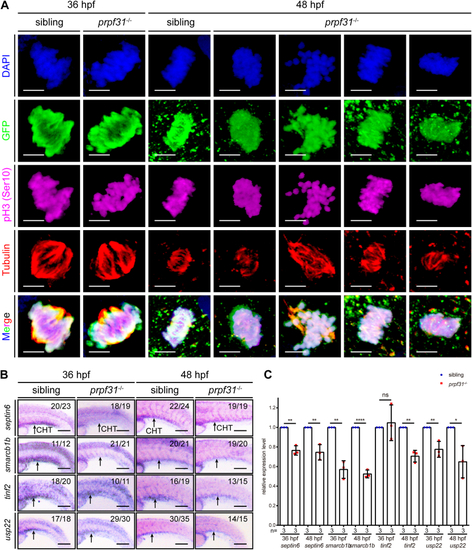
Aberrant alternative splicing of mitosis-related genes leads to mitotic malformations of HSPCs in the CHT of prpf31−/−zebrafish.A, quadruple staining of DAPI, cmyb:EGFP, pH3 (Ser10), and tubulin showed the mitotic status of HSPCs in the CHT of prpf31−/− zebrafish and siblings at 36 and 48 hpf. At least 25 mitotic HSPCs in more than six embryos were observed for each group. The scale bars represent 4 μm. B, expression of four aberrantly spliced mitosis-related genes (septin6, smarcb1b, tinf2, and usp22) in the CHT of siblings and prpf31−/− zebrafish at 36 and 48 hpf by WISH. Lateral views. Black arrows denote the CHT region. The number of embryos with similar gene expression patterns among all embryos examined were shown on the top right of each panel. The scale bars represent 100 μm. C, qRT-PCR analysis of four aberrantly spliced mitosis-related genes (septin6, smarcb1b, tinf2, and usp22) in siblings and prpf31−/− zebrafish at 36 and 48 hpf. Data were shown as mean ± SD of three independent experiments (n = 3); unpaired two-tailed t test; ∗p < 0.05, ∗∗p < 0.01, ∗∗∗∗p < 0.0001; ns, not significant. CHT, caudal hematopoietic tissue; hpf, hours postfertilization; HSPC, hematopoietic stem and progenitor cell; pH3, phospho-histone 3; qRT-PCR, quantitative real-time PCR; WISH, whole-mount in situ hybridization. DAPI, 4′,6-diamidino-2-phenylindole; EGFP, enhanced green fluorescent protein.

Model of Prpf31 action in HSPC expansion during zebrafish embryogenesis. In zebrafish embryogenesis, the nascent HSPCs undergo extensive proliferation for pool expansion and commitment-proliferation-differentiation in the CHT, which demand rapid and periodic regulated pre-mRNA alterative splicing. Accurate and sequential regulated pre-mRNA alterative splicing ensure efficient and precise HSPCs mitosis for rapid blood replenishment. In prpf31−/− zebrafish, the spliceosomes cannot assemble effectively due to deficiency of Prpf31, which compromises the splicing efficiency of the spliceosome machine, and results in aberrant mRNA alternative splicing, perturbs the alternative splicing of mitosis-related genes, predisposes HSPCs to malformed mitosis and cell cycle arrest in M phase, and eventually impaired the expansion and differentiation of HSPCs in the CHT. CHT, caudal hematopoietic tissue; HSPC, hematopoietic stem and progenitor cell; PRPF, pre-mRNA processing factor.

Acknowledgments
This image is the copyrighted work of the attributed author or publisher, and ZFIN has permission only to display this image to its users. Additional permissions should be obtained from the applicable author or publisher of the image. Full text @ J. Biol. Chem.