FIGURE SUMMARY
Title

Apela promotes blood vessel regeneration and remodeling in zebrafish

Authors
Nys, N., Khatib, A.M., Siegfried, G.
Source
Full text @ Sci. Rep.

Apela is upregulated during zebrafish caudal fin regeneration. (A) Schematic representation of experimental plan showing amputation site and regenerated fin areas. Adult zebrafish were anesthetized in tricaine and fins were amputated and allowed to regenerate for indicated time periods. (B) Total RNA was isolated from fins (15–20 fins per time point) with uncut fins as controls (0 dpa) and analyzed by real-time PCR using specific primers for Apela or β-actin. Results are shown in the bar graph and are expressed as the ratio of the indicated transcripts relative to control (0 dpa). Results are shown as means ± S.E. of three experiments performed in triplicate. (C) Representative confocal images of control fins (0 dpa) and at 3 dpa (n = 12) subjected to immunofluorescence of Apela (red signal). (D) Quantification of fluorescence intensity in ImageJ. (E) Total RNA was isolated from fins (15–20 fins per time point) with uncut fins as controls (0 dpa) and analyzed by real-time PCR as in (A), using specific primers for Apln. (F) Aplnra and Aplnrb expression analysis by RT-PCR in uncut fins. (G) Aplnra expression analysis as in (A). The mean ± S.E values are shown. *p < 0.05. Scale bars represent 0.5 mm.

Blockade of Apela expression suppresses vessel differentiation. (A, B) Representative confocal images of caudal fins of adult zebrafish (15–20 fins/per experiment) that were electroporated with control Mo-CTL and Mo directed against Apela (Mo-Apela), and fin were allowed to regenerate for 3 days prior Apela expression analysis by immunohistochemistry using an anti-Apela. (C, D) MO-Apela had no effect on total fin regeneration. (E) Schematic representation of anastomotic bridge with emerging sprouts forming vascular plexus and the U-shaped formed by the artery and the two veins. (F) Inhibition of Apela by MO-Apela prevents remodeling of immature vascular network during fin regenerating in Tg(fli:EGFP)y, as observed with the enhanced plexus (G) and branching (H) vasculature. Results are shown as means ± S.E. of three experiments performed in triplicate. *p < 0.05. NS, not significant.

Apela mediates fin vessel differentiation during regeneration. (A, B) Tg(fli:EGFP)y1 zebrafish exposed to Apela (10 μM) after fin amputation and were allowed to regenerate prior analysis. (C, E) Tg(fli:EGFP)y1 fins (20 fins/per experiment) treatment with Apela (10 μM) shows reduced vasculature plexus (D) and branching (E). (F, G) Western blot analysis of the activation of AKT in control fins and after 15 min treatment with Apela (10 μM). Images shown in upper and lower panels were cropped from the same blot. Full blots are provided in Supplementary Fig. 1. All results shown are representative of at least 3 independent experiments with means ± S.E and *p < 0.05.

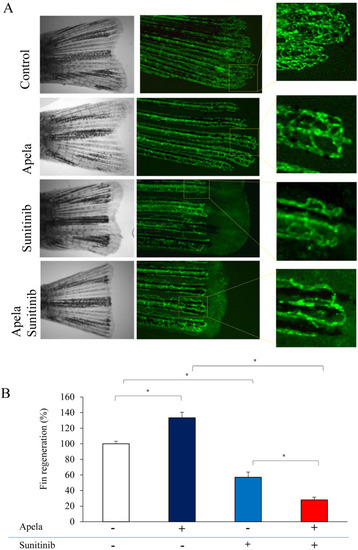
Sunitinib and Apela cross talk during vascular plexus regeneration and remodeling. (A) Zebrafish (15–20/per experiment) exposed to Sunitinib (0.5 μM) in the absence and presence of Apela (10 μM) after fin amputation and allowed to regenerate during 3 days prior analysis. (B) Results are shown as means ± S.E. of three experiments performed in triplicate. *p < 0.05.

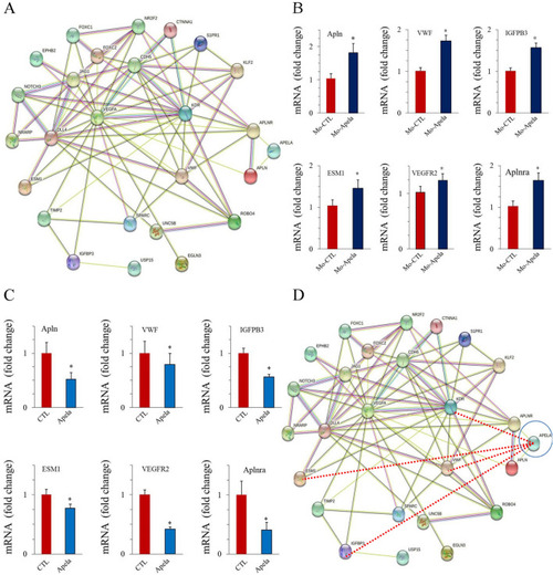
Apela and vascular plexus remodeling molecule network. (A) The STRING network view. Combined screenshots from the STRING website, which has been queried with a subset of proteins involved in the formation and remodeling of vascular vessels (Notch, VEGF, Ephrin, Hedgehog, TGF, COUP-TFII, FOXC2 and others. Colored lines between the proteins indicate the interaction evidence. (B, C) Relative expression of indicated VWF, IGFPB3, ESM1, VEGFR2, Apela, Apln and Aplnr mRNA in pooled fin zebrafish (15–20/per experiment) treated with Apela (B) or Mo-Apela (C). (D) STRING network queried with the same subset of proteins in (A) and the set of the angiogenic proteins: VWF, IGFPB3, ESM1, VEGFR2, Apela, Apln and Aplnr. Red lines between the proteins indicate the new interaction evidence. For (B) and (C), results are shown as means ± S.E. of three experiments performed in triplicate. *p < 0.05.

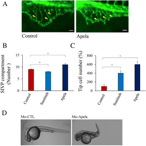
Effect of Apela on sub-intestinal venous plexus (SIVP) formation. (AC) Tg(fli:EGFP)y1 embryos zebrafish (15–20/per experiment) were treated with Apela and sunitinib (for comparison) and the number of SIV compartments (B) and tip cells (C) were enumerated. Yellow arrowheads point to compartments. (D) Injection of MO-Apela into zebrafish embryos at the 1–4 cell stage, significantly affected normal embryonic development. For (B) and (C), results are shown as means ± S.E. of three experiments performed in triplicate. *p < 0.05.

Acknowledgments
This image is the copyrighted work of the attributed author or publisher, and ZFIN has permission only to display this image to its users. Additional permissions should be obtained from the applicable author or publisher of the image. Full text @ Sci. Rep.