IMAGE

Figure 6

ID
ZDB-IMAGE-240217-6
Source
Figures for Nys et al., 2024
Image
Figure Caption

Figure 6

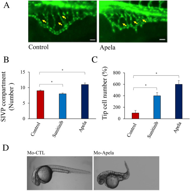
Effect of Apela on sub-intestinal venous plexus (SIVP) formation. (AC) Tg(fli:EGFP)y1 embryos zebrafish (15–20/per experiment) were treated with Apela and sunitinib (for comparison) and the number of SIV compartments (B) and tip cells (C) were enumerated. Yellow arrowheads point to compartments. (D) Injection of MO-Apela into zebrafish embryos at the 1–4 cell stage, significantly affected normal embryonic development. For (B) and (C), results are shown as means ± S.E. of three experiments performed in triplicate. *p < 0.05.

Acknowledgments
This image is the copyrighted work of the attributed author or publisher, and ZFIN has permission only to display this image to its users. Additional permissions should be obtained from the applicable author or publisher of the image. Full text @ Sci. Rep.