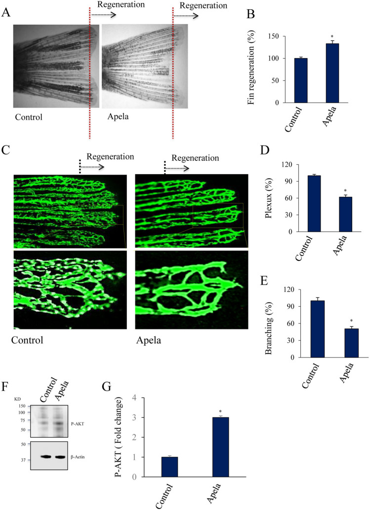
Apela mediates fin vessel differentiation during regeneration. (A, B) Tg(fli:EGFP)y1 zebrafish exposed to Apela (10 μM) after fin amputation and were allowed to regenerate prior analysis. (C, E) Tg(fli:EGFP)y1 fins (20 fins/per experiment) treatment with Apela (10 μM) shows reduced vasculature plexus (D) and branching (E). (F, G) Western blot analysis of the activation of AKT in control fins and after 15 min treatment with Apela (10 μM). Images shown in upper and lower panels were cropped from the same blot. Full blots are provided in Supplementary Fig. 1. All results shown are representative of at least 3 independent experiments with means ± S.E and *p < 0.05.
Acknowledgments
This image is the copyrighted work of the attributed author or publisher, and
ZFIN has permission only to display this image to its users.
Additional permissions should be obtained from the applicable author or publisher of the image.
Full text @ Sci. Rep.
Your Input Welcome
Thank you for submitting comments. Your input has been emailed to ZFIN curators who may contact you if
additional information is required.
Oops. Something went wrong. Please try again later.