FIGURE SUMMARY
Title

A Novel Anticancer Peptide Derived from Bryopsis plumosa Regulates Proliferation and Invasion in Non-Small Cell Lung Cancer Cells

Authors
Kim, H., Kim, H.T., Jung, S.H., Han, J.W., Jo, S., Kim, I.G., Kim, R.K., Kahm, Y.J., Choi, T.I., Kim, C.H., Lee, J.H.
Source
Full text @ Mar. Drugs

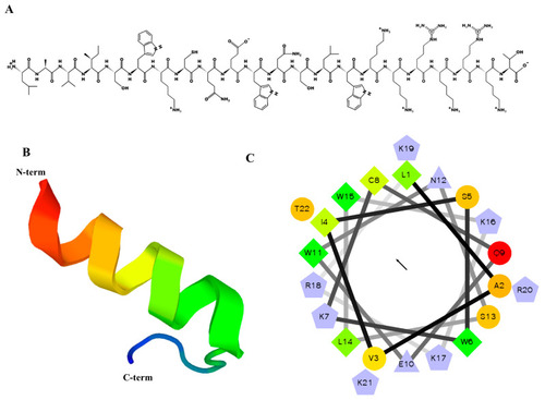
Predicted helical secondary and three-dimensional structures of MP06 anticancer peptide. (A) Two-dimensional drawing of the peptide using the PepDraw software (https://pepdraw.com, accessed on 12 April 2022) (B) The predicted three-dimensional secondary structure of MP06 from PEP-FOLD3. (C) Helical wheel plots of MP06 sequences showing amphipathicity quantified as hydrophobic moments. Shows hydrophilic residues as circles, hydrophobic residues as diamonds, and potentially positively/negatively charged as triangles/pentagons. The most hydrophobic residue is green, and the amount of green decreases proportionally to the hydrophobicity, with zero hydrophobicity coded as yellow. Hydrophilic residues are coded red, with pure red being the most hydrophilic (uncharged) residue and the amount of red decreasing proportionally to the hydrophilicity. The potentially charged residues are light blue. The arrow in the middle of the circle indicates the hydrophobic direction formed by amino acids with high hydrophobicity.

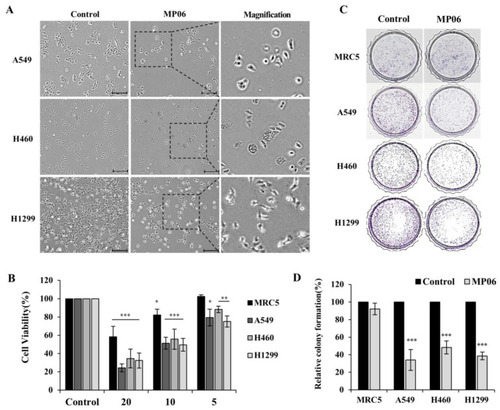
Investigating the role of MP06 anticancer peptide in NSCLCs. (A) Morphologic changes of A549, H460 and H1299 cells treated with 10 μM MP06 compared water-treated control cells. (B) Effect of MP06 at a concentration of 20, 10 and 5 μM on the growth of normal lung fibroblast and NSCLCs for 48 h. (C) The difference in colony forming ability on treatment of 10 μM MP06 in lung cells. (D) Quantification of relative colony formation in NSCLCs treated with MP06. Scale bars: 200 µm. * p < 0.05, ** p < 0.01 and *** p < 0.001.

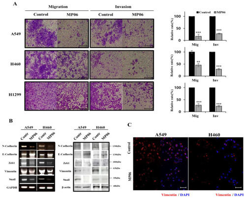
Regulation of epithelial–mesenchymal transition (EMT) in NSCLCs by MP06. (A) Influence MP06 on NSCLCs migration (24 h) and invasion (48 h). Scale bars: 200 µm. (B) Expression level of epithelial–mesenchymal transition (EMT) markers with 10 μM MP06 compared water-treated control cells (contr) in NSCLCs for 48 h using RT-PCR and Western blotting assay. GAPDH and β-actin were used as housekeeping genes on the basis of their consistency of expression. (C) The expression of vimentin in A549 and H460 cells treated with MP06 using Immunofluorescence (IF) analysis. Scale bars: 100 µm. ** p < 0.01 and *** p < 0.001.

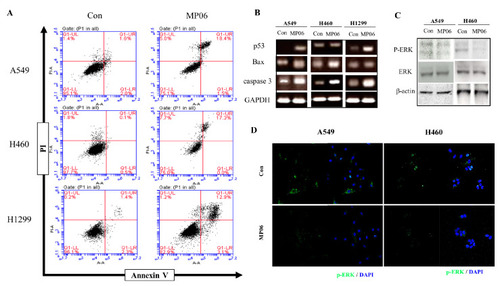
The effect of MP06 on apoptosis of NSCLCs. (A) Detection of apoptosis and necrosis in NSCLCs via Annexin V/PI apoptosis kit staining. (B) Expression of apoptosis-related genes such as P53, Bax, and caspase3 in NSCLCs treated with 10 μM MP06. (C) The expression of phosphorylation ERK in MP06 treated A549 and H460 cells using Western blotting assay. (D) The expression of p-ERK in A549 and H460 cells treated with MP06 using IF analysis. Scale bars: 100 µm.

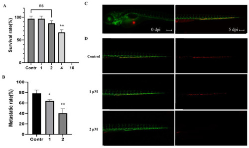
Inhibition of metastasis on A549 cells with MP06 in zebrafish embryos (A) Survival evolution through time of 48 h post fertilization (hpf) zebrafish embryos exposed to MP06 at different concentrations. (B) Quantification of metastatic rate of Tg(kdrl:GFP) zebrafish embryos microinjected with CM-DiI A549 cells at 5 dpi in zebrafish xenograft treated with MP06 (C) Injected A549 cells (red) immediately after xenograft (left) and at 5 dpi (right) (D) Xenografted zebrafish treated with 1, 2 μM MP06. Representative images show the invasive A549 cells (red) in the tail region of the embryos via vessels (green). (n = 30, ns: not statistically significant, Scale bars: 200 µm. * p < 0.01, ** p < 0.001).

Acknowledgments
This image is the copyrighted work of the attributed author or publisher, and ZFIN has permission only to display this image to its users. Additional permissions should be obtained from the applicable author or publisher of the image. Full text @ Mar. Drugs