IMAGE

Figure 5

ID
ZDB-IMAGE-231225-29
Source
Figures for Kim et al., 2023
Image
Figure Caption

Figure 5

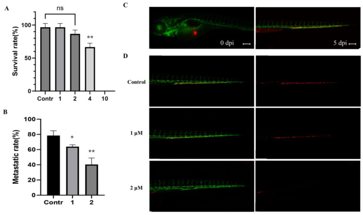
Inhibition of metastasis on A549 cells with MP06 in zebrafish embryos (A) Survival evolution through time of 48 h post fertilization (hpf) zebrafish embryos exposed to MP06 at different concentrations. (B) Quantification of metastatic rate of Tg(kdrl:GFP) zebrafish embryos microinjected with CM-DiI A549 cells at 5 dpi in zebrafish xenograft treated with MP06 (C) Injected A549 cells (red) immediately after xenograft (left) and at 5 dpi (right) (D) Xenografted zebrafish treated with 1, 2 μM MP06. Representative images show the invasive A549 cells (red) in the tail region of the embryos via vessels (green). (n = 30, ns: not statistically significant, Scale bars: 200 µm. * p < 0.01, ** p < 0.001).

Acknowledgments
This image is the copyrighted work of the attributed author or publisher, and ZFIN has permission only to display this image to its users. Additional permissions should be obtained from the applicable author or publisher of the image. Full text @ Mar. Drugs