FIGURE SUMMARY
Title

Bi-allelic variants in FLII cause pediatric cardiomyopathy by disrupting cardiomyocyte cell adhesion and myofibril organization

Authors
Ruijmbeek, C.W., Housley, F., Idrees, H., Housley, M.P., Pestel, J., Keller, L., Lai, J.K.H., van der Linde, H.C., Willemsen, R., Piesker, J., Al-Hassnan, Z.N., Almesned, A., Dalinghaus, M., van den Bersselaar, L.M., van Slegtenhorst, M.A., Tessadori, F., Bakkers, J., van Ham, T.J., Stainier, D.Y., Verhagen, J.M., Reischauer, S.
Source
Full text @ JCI Insight

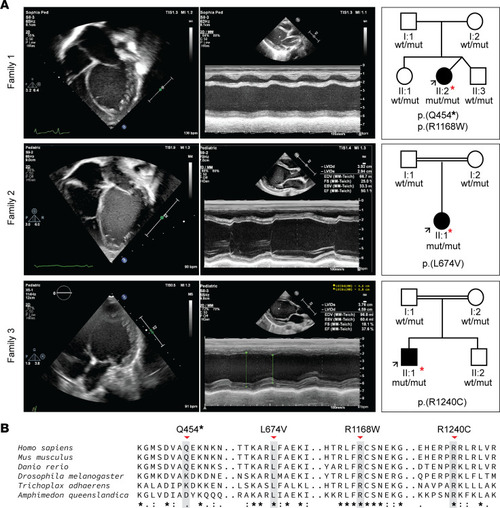
Clinical manifestation of early-onset DCM and family pedigree of affected individuals included in this study.

(A) Echo findings at presentation. Left panel, per family: 2-dimensional, apical 4-chamber echocardiographic image of the probands depicting an enlarged and spherically shaped left ventricle. Middle panel: M-mode echocardiography displaying severely depressed left ventricular (LV) function. Right panel: family pedigrees that were found to segregate biallelic variants in the FLII gene. I and II refer to the first and second generations of the family, respectively. The arrow points to the proband. (B) Alignment of FLII protein sequence across the metazoan kingdom including orthologs from invertebrates and vertebrates. Note the full conservation of the affected amino acid residues and high contextual conservation in a wide range of species ranging from simple multicellular organisms including sponges (Amphimedon queenslandica) and placozoa (Trichoplax adhaerens) to higher species including insects (Drosophila melanogaster), bony fishes (Danio rerio), rodents (Mus musculus), and primates (Homo sapiens) illustrating functional significance.

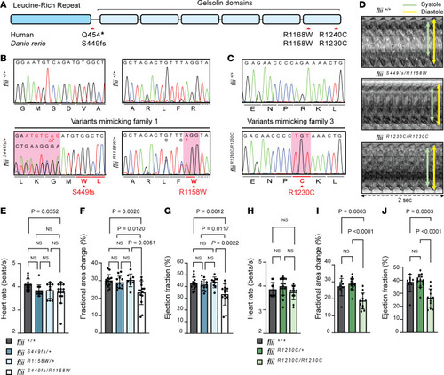
CRISPR/Cas9-mediated genome editing of patient-specific biallelic variants in flii in zebrafish results in DCM-associated phenotypes in early development.

(A) Schematic representation of the FLII protein, consisting of a leucine-rich repeat (LRR) and 6 gelsolin-like domains. Location of the human variants of families 1 and 3 are depicted with corresponding variants generated in zebrafish (D. rerio). (B) Aligned Sanger sequencing traces of wild-type flii+/+ and genome-edited PCR amplicons from compound heterozygous fliiS449fs/R1158W larvae harboring a heterozygous variant resulting in a frameshift starting at amino acid position 449 (left panel, sequence is reverse complement as it was sequenced with the reverse primer) and the heterozygous R1158W missense variant (right panel), representing family 1. Modified codons are underlined in red. Dotted line represents intronic sequence. (C) Representative Sanger sequencing results of the PCR amplicons from wild-type and fliiR1230C/R1230C zebrafish larvae harboring the homozygous R1230C missense variant, representing family 3. The modified codon is underlined in red. (D) Ventricular kymographs derived from high-speed imaging video recordings of 120 hpf zebrafish larvae spanning approximately 2 seconds: flii+/+ (top panel), fliiS449fs/R1158W (middle panel) and fliiR1230C/R1230C (lower panel). Note that there are no signs of irregular heart rhythm in larvae harboring patient-specific biallelic variants. (EG) Ventricular contractility parameters derived from high-speed imaging movies, including heart rate (E), fractional area change (F), and ejection fraction (G) for flii+/+, fliiS449fs/+, fliiR1158W/+, and fliiS449fs/R1158W. flii+/+n = 14; fliiS449fs/+n = 12; fliiR1158W/+n = 9; fliiS449fs/R1158Wn = 14. (HJ) Ventricular contractility parameters derived from high-speed imaging movies, including heart rate (H), fractional area change (I), and ejection fraction (J) for flii+/+, fliiR1230C/+, and fliiR1230C/R1230C. flii+/+n = 9; fliiR1230C/+n = 15; fliiR1230C/R1230Cn = 12. Statistics: mean ± SD; 1-way ANOVA coupled with Tukey’s multiple-comparison test was used to test for significance.

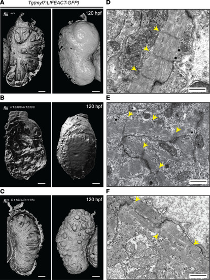
Flii dysfunction results in myofibrillar architectural abnormalities of the ventricular myocardium.

(AC) 3D volume renderings of maximum projections of Tg(myl7:LIFEACT-GFP) cardiac ventricles at 120 hpf from wild-type flii+/+ (A), patient-specific fliiR1230C/R1230C (B), and fliiD110fs/D110fs (C); left panels show ventricular lumen; right panels show ventricular surface. Note that the complex trabecular network observed in wild-type is affected in both mutant alleles. In the severe loss-of-function fliiD110fs mutants, some of the epithelial shaped cardiomyocytes adopt a spherical shape and blebb out of the ventricular wall. Scale bars: 50 μm. Sample size for each genotype, n ≥ 3 biological replicates. (DF) Representative TEM images of ventricular cardiac muscle from 120 hpf larvae, showing well-organized bundled myofibrils and z-discs in wild-type flii+/+ (D), which are disorganized in patient-specific fliiR1230C/R1230C mutants (E) and appear to be more severely affected in fliiD110fs/D110fs mutants with faintly present z-discs (F). Yellow arrows, z-discs. Scale bars, 1 μm. Sample size for each genotype, n ≥ 3 biological replicates.

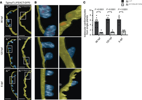
Loss of Flii results in a reduced number of cardiac trabeculae.

(A) Single confocal planes of representative Tg(myl7:LIFEACT-GFP) ventricular outer curvatures at 96 hpf through 6 dpf of flii+/? siblings (left) and fliiD110fs/D110fs mutants (right). The 96 hpf hearts express Tg(myl7:nDsRed2) to visualize nuclei. Yellow false color depicts outer curvature. Trabecular myocardium is highlighted in blue. Scale bars: 25 μm. (B) Cropped areas corresponding to white rectangles in A. The dotted lines depict the compact layer of cardiomyocytes. (C) Quantification of protruding cardiomyocytes out of the single-layered compact myocardium in the ventricular outer curvature from sagittal planes at 96 hpf through 6 dpf. Unpaired t test; values represent means ± SEM; (96 hpf flii+/? siblings n = 6, fliiD110fs/D110fsn = 9); (120 hpf flii+/? siblings n = 5, fliiD110fs/D110fsn = 5); (6 dpf flii+/? siblings n = 3, fliiD110fs/D110fsn = 3).

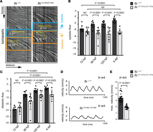
Blood flow analysis reveals reduced cardiac performance upon Flii deficiency, including a developmental arrest in the systolic hemodynamic force.

(AC) Analysis of blood flow velocity (BFV) in the dorsal aorta by spinning disk microscopy at 72 hpf through 6 dpf. (A) Blood flow videos (400 frames/s, total of 500 frames shown) are visualized as kymographs, which show dynamics of blood cells that move distance x over frames y. Relative speeds are determined by measuring the angle of blood flow in the kymographs, with a steeper downward angle representing slower blood flow. In the systolic phase (blue box), blood cells move faster than in the diastolic phase (orange box). (B and C) Quantification of kymograph angles in systolic and diastolic phases, respectively. Note that the systolic blood cell speed does not increase in fliiD110fs mutants with developmental time (B). In contrast, the diastolic blood cell speed increases in fliiD110fs mutants but is still significantly reduced compared with that of wild-type and heterozygous siblings (C). One-way ANOVA coupled with Holm-Šídák multiple-comparison test was used to test for significance; values represent means ± SEM; (72 hpf flii+/? siblings n = 7, fliiD110fs/D110fsn = 9); (96 hpf flii+/? siblings n = 9, fliiD110fs/D110fsn = 8); (120 hpf flii+/? siblings n = 9, fliiD110fs/D110fsn = 8); (6 dpf flii+/? siblings n = 8, fliiD110fs/D110fsn = 6). (D and E) Quantification of absolute blood cell speed by single-cell tracking at 6 dpf reveals a normal sinus rhythm of heartbeats in both fliiD110fs/D110fs and flii+/? siblings. Bar graphs display maximum velocity of blood cells in the dorsal aorta. Unpaired t test; values represent means ± SEM.

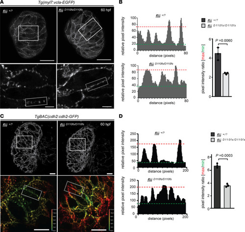
Flii-deficient zebrafish exhibit defects in vinculin-EGFP and cadherin2-EGFP localization.

(A) 3D confocal projections of 60 hpf Tg(myl7:vcla-EGFP) flii+/? sibling and fliiD110fs/D110fs cardiac ventricles. Vinculin-EGFP expression is restricted to the lateral membranes. Note that vinculin-EGFP expression is concentrated into foci in siblings but appears more diffuse in fliiD110fs/D110fs zebrafish (magnifications shown in lower panel); each group, n = 5. Scale bars: projections, 25 μm; magnifications, 5 μm. (B) Plots of the relative pixel intensity along membranes from dotted boxed areas of A. Green and red dotted lines correspond to average minimum and maximum relative pixel intensities, respectively. Quantification of pixel intensity ratios is shown on the right. Unpaired t test; values represent means ± SEM; each group, n = 3. (C) Representative 3D views of 60 hpf TgBAC(cdh2:cdh2-EGFP)flii+/? sibling (left panels) and fliiD110fs/D110fs cardiac ventricles (right panels). Magnifications show a clear punctate localization of cadherin2-EGFP in wild-type controls that is lacking in fliiD110fs/D110fs embryos (Z-plane position color coded as indicated); each group n = 5. Scale bars: projections, 10 μm; magnifications, 10 μm. (D) Plots of the relative pixel intensity along membranes from dotted boxed areas of C. Green and red dotted lines correspond to average minimum and maximum relative pixel intensities, respectively. Quantification of pixel intensity ratios is shown on the right. Unpaired t test; values represent means ± SEM; n = 3 for each genotype.

Aberrant activation of the Notch and Hippo signaling pathways in Flii-deficient ventricles.

(A) Representative 3D volume renderings of TP1bglob:VenusPEST of flii+/? sibling and fliiD110fs/D110fs hearts at 96 hpf. White dotted line outlines the heart. Note that there is no Notch reporter expression in the ventricle of fliiD110fs/D110fs hearts, whereas there is expression in their AVC and OFT. Each group n = 5. V, ventricle; A, atrium; AVC, atrioventricular canal; OFT, outflow tract. (B) Representative maximum-intensity projections of wholemount flii+/? sibling and fliiD110fs/D110fs hearts at 60 hpf stained for Wwtr1. flii+/? siblings n = 7, fliiD110fs/D110fsn = 11. (C) Corresponding confocal sagittal sections of the wholemount ventricles shown in B. Nuclei are counterstained with DAPI, and cardiomyocyte F-actin myofibrils are marked with myl7:LIFEACT-GFP expression; scale bars, 20 μm.

Acknowledgments
This image is the copyrighted work of the attributed author or publisher, and ZFIN has permission only to display this image to its users. Additional permissions should be obtained from the applicable author or publisher of the image. Full text @ JCI Insight