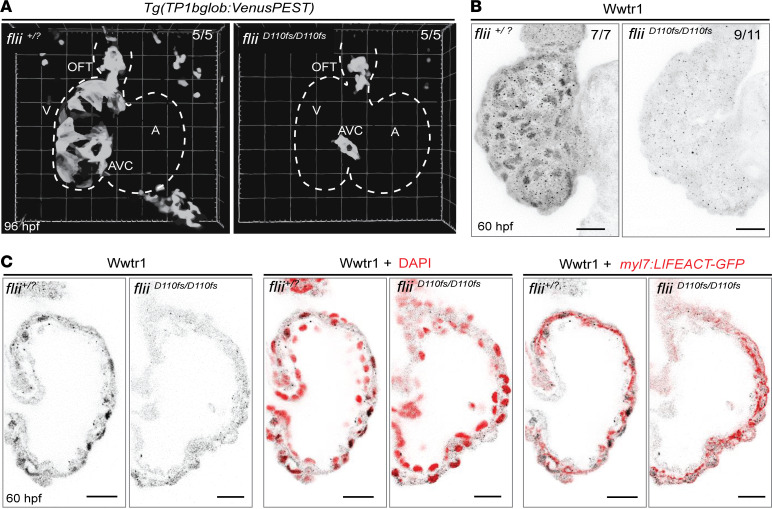
Figure 7
Aberrant activation of the Notch and Hippo signaling pathways in Flii-deficient ventricles.
(A) Representative 3D volume renderings of TP1bglob:VenusPEST of flii+/? sibling and fliiD110fs/D110fs hearts at 96 hpf. White dotted line outlines the heart. Note that there is no Notch reporter expression in the ventricle of fliiD110fs/D110fs hearts, whereas there is expression in their AVC and OFT. Each group n = 5. V, ventricle; A, atrium; AVC, atrioventricular canal; OFT, outflow tract. (B) Representative maximum-intensity projections of wholemount flii+/? sibling and fliiD110fs/D110fs hearts at 60 hpf stained for Wwtr1. flii+/? siblings n = 7, fliiD110fs/D110fsn = 11. (C) Corresponding confocal sagittal sections of the wholemount ventricles shown in B. Nuclei are counterstained with DAPI, and cardiomyocyte F-actin myofibrils are marked with myl7:LIFEACT-GFP expression; scale bars, 20 μm.
Acknowledgments
This image is the copyrighted work of the attributed author or publisher, and
ZFIN has permission only to display this image to its users.
Additional permissions should be obtained from the applicable author or publisher of the image.
Full text @ JCI Insight
Your Input Welcome
Thank you for submitting comments. Your input has been emailed to ZFIN curators who may contact you if
additional information is required.
Oops. Something went wrong. Please try again later.