
- Title
-
Identification of an α-l-iduronidase (IDUA) M1T mutation in a Chinese family with autosomal recessive mucopolysaccharidosis I
- Authors
- Liu, D., Jiang, Z., Deng, L., Li, H., Jiang, H.
- Source
- Full text @ Ann N Y Acad Sci
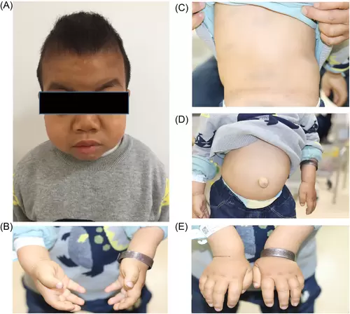
Patient's appearance. (A) Anthropometric examination showed the patient was severely stunted and presented with coarse facial features. (B) Claw-like appearance of both hands. (C) Scoliosis and kyphosis deformity of spine. (D) Umbilical hernia. (E) Claw-like hands |
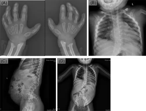
X-ray of hands, chest, and abdomen showed dysostosis multiplex. (A) X-ray of both hands showed widened phalanges and metacarpals with proximal pointing. (B) X-ray of chest showed the gross cardiomegaly with widened anterior end of ribs. (C, D) X-ray of abdomen showed deformation of external clavicle, bone defect of anterior edge of L1 vertebral body, multiple vertebral body morphological disorders, and some rib morphological disorders (e.g., ribbon-like changes). |
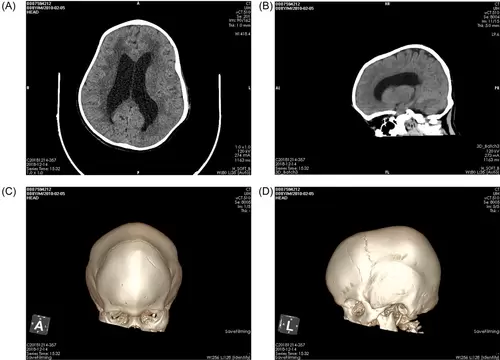
Head CT showed dolichocephalic head with (A) ventricular enlargement, (B) hydrocephalus, and (C, D) premature closure of the herringbone suture. Abbreviations: A, anterior; CT, computed tomography; L, left |
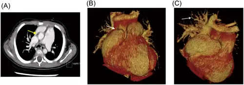
Cardiac CT with contrast demonstrated enlargement of right atrium and ventricle, pulmonary hypertension, and partial anomalous pulmonary venous connection. (A) The axial image demonstrated obviously dilated pulmonary trunk, which was almost twice of the aorta (yellow arrow). The superior vena cava was also dilated as entering the right atrium, receiving anomalous drainage of right upper and middle pulmonary vein drainage. (B) The frontal view of volume reformatted image demonstrated an enlarged heart, especially the right atrium (white arrow). The prominent right atrium auricle was just below the left brachiocephalic vein. (C) The posterior view showed the anomalous pulmonary veins (white arrow), which were distinct from the other side. Abbreviation: CT, computed tomography. |
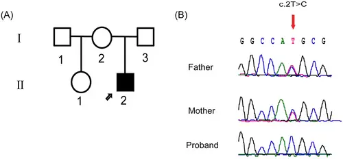
Mutation analysis of IDUA in this pedigree. (A) Pedigree of the family. Proband is noted with black arrow. (B) DNA sequence chromatogram (forward strand) shows a homozygous T>C transition that causes a substitution of threonine for methionine at codon 1. The red arrow points to the position of the mutant nucleotide. |
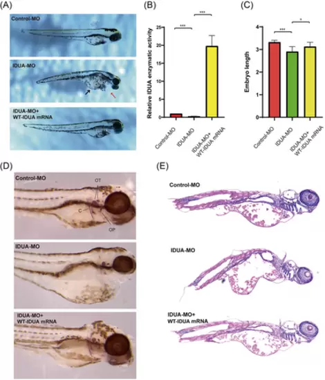
Depletion of IDUA in zebrafish results in multiple developmental defects. (A) Representative stereomicroscopy images of control-MO, IDUA-MO, and IDUA-MO + WT-IDUA mRNA zebrafish larvae at 3 dpf. IDUA-MO zebrafish larvae had protracted jaws (red arrow), enlarged hearts, and pericardial edema (black arrow). Introduction of WT-IDUA mRNA in the morphant zebrafish fully rescued these phenotypes. (B) The enzymatic activities of IDUA in each group were assayed. IDUA enzymatic activity in the control group was normalized to 1, the enzyme activity from IDUA-MO and IDUA-MO + WT-IDUA mRNA zebrafish embryos was presented as fold induction above this level. Note: Data are representative of three individual experiments. (C) Quantitative analysis of the whole-body length of each group. As measured in millimeters (n = 15 per condition; Student's t-test; ***p<0.001; data are representative of three individual experiments). (D) Alizarin red bone staining of 5 dpf control-MO, IDUA-MO, and IDUA-MO + WT-IDUA mRNA zebrafish larvae. (E) Sagittal section images of hematoxylin and eosin-stained tissue from 5 dpf control-MO, IDUA-MO, and IDUA-MO + WT-IDUA mRNA zebrafish larvae. Abbreviations: C, cleithrum; IDUA, α-l-iduronidase; MO, morpholino; OP, operculum; OT, otoliths; WT, wild type. Student's t-test; *p<0.05, ***p<0.001. |

Increased apoptosis in IDUA-deficient zebrafish larvae. TUNEL staining of cryopreserved tissue sections from control-MO, IDUA-MO, and IDUA-MO + WT-IDUA mRNA zebrafish larvae at 5 dpf. TUNEL-positive cells were detected in the eye, brain, and internal organs of IDUA-MO zebrafish larvae. Cell nuclei are stained with DAPI (blue). TUNEL image (first column), DAPI images (second column), and merged TUNEL/DAPI images (third column) are shown. All the images are representative of at least three independent experiments. Abbreviations: IDUA, α-l-iduronidase; MO, morpholino |
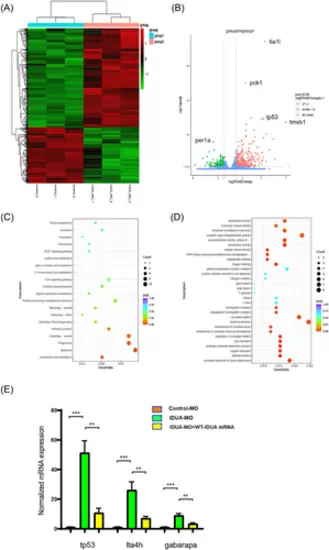
RNA sequencing analyses reveal differentially expressed genes and pathways in response to knockdown of IDUA in zebrafish larvae. (A) The heat map clustering of 3 dpf control-MO and IDUA-MO zebrafish larvae. Red blocks represent the upregulated genes and the green blocks represent downregulated genes. (B) Volcano plot of the genes with significantly different expression in 3 dpf control-MO and IDUA-MO zebrafish larvae. (C) KEGG analysis of the differentially expressed genes. (D) GO biological processes analysis of the differentially expressed genes. (E) Expression of tp53, lta4h, and gabarapa mRNA was detected by qPCR in 3 dpf control-MO, IDUA-MO, and IDUA-MO + WT-IDUA mRNA zebrafish larvae. **p<0.01, ***p<0.001. |