
Fig. 4
- ID
- ZDB-FIG-231212-72
- Publication
- Liu et al., 2023 - Identification of an α-l-iduronidase (IDUA) M1T mutation in a Chinese family with autosomal recessive mucopolysaccharidosis I
- Other Figures
- All Figure Page
- Back to All Figure Page
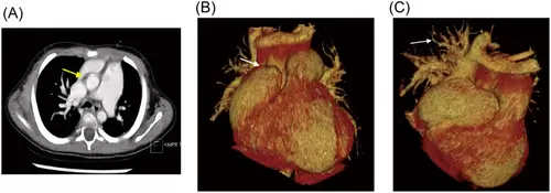
Cardiac CT with contrast demonstrated enlargement of right atrium and ventricle, pulmonary hypertension, and partial anomalous pulmonary venous connection. (A) The axial image demonstrated obviously dilated pulmonary trunk, which was almost twice of the aorta (yellow arrow). The superior vena cava was also dilated as entering the right atrium, receiving anomalous drainage of right upper and middle pulmonary vein drainage. (B) The frontal view of volume reformatted image demonstrated an enlarged heart, especially the right atrium (white arrow). The prominent right atrium auricle was just below the left brachiocephalic vein. (C) The posterior view showed the anomalous pulmonary veins (white arrow), which were distinct from the other side. Abbreviation: CT, computed tomography. |