
- Title
-
Holoprosencephaly with a Special Form of Anophthalmia Result from Experimental Induction of bmp4, Oversaturating BMP Antagonists in Zebrafish
- Authors
- Bulk, J., Kyrychenko, V., Rensinghoff, P.M., Ghaderi Ardekani, Z., Heermann, S.
- Source
- Full text @ Int. J. Mol. Sci.
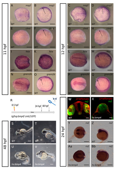
(A–Q): expression of bmp-antagonists nog2, chrd, fsta and grem2b in the ANP at 11 and 12 hpf. (A–D): ISH for nog2. (A,B): 11 hpf; (C,D): 12 hpf. (E–H): ISH for chrd. (E,F): 11 hpf; (G,H): 12 hpf. (I–M): ISH for fsta. (I,K): 11 hpf; (L,M): 12 hpf. (N–Q): ISH for grem2b. (N,O): 11 hpf; (P,Q): 12 hpf. (A,C,E,G,I,L,N,P): dorsal view. (B,D,F,H,K,M,O,Q): lateral view. Scalebars indicate 200 µm. (R): summary of experimental procedure. (S–Bb): Embryos were heat shocked at 8.5 hpf and observed at 24 hpf or 48 hpf, resp. (S,T): bright-field image of wildtype at 48 hpf. (U,V): bmp4-induced larva at 48 hpf. (S,U): dorsal view. (T,V): lateral view. Scalebars indicate 250 µm. (W,X): transversal confocal sections of larvae with rx2:GFP at 24 hpf. injection of LY-tdTomato mRNA in zygote. (W): wildtype. (X): bmp4-induced, scalebars indicate 50 µm. (Y–Bb): ISH for rx2 at 24 hpf. (Y,Z): wildtype. (Aa,Bb): bmp4-induced. (Y,Aa): dorsal view; (Z,Bb): lateral view. Scalebars indicate 250 µm. |
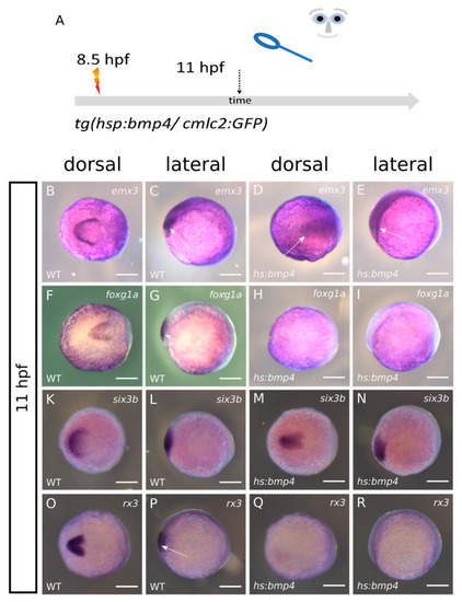
ISH at 11 hpf after bmp4-induction at 8.5 hpf. left columns wildtypes, right columns bmp4-induced. first and third column dorsal view, second and fourth column lateral view. (A): summary of experimental procedure. Embryos were heat-shocked at 8.5 hpf and analyzed at 11 hpf. (B–E): Expression of emx3 is condensed at the midline after bmp4-induction (arrows). (F–I): the foxg1a domain in the forebrain (arrow) is lost after bmp4-induction. (K–N): expression of eye-field marker six3b is also condensed at the midline after bmp4-induction. (O–R): expression of eye-field marker rx3 (arrow) is lost after bmp4-induction. Scalebars indicate 200 µm. |
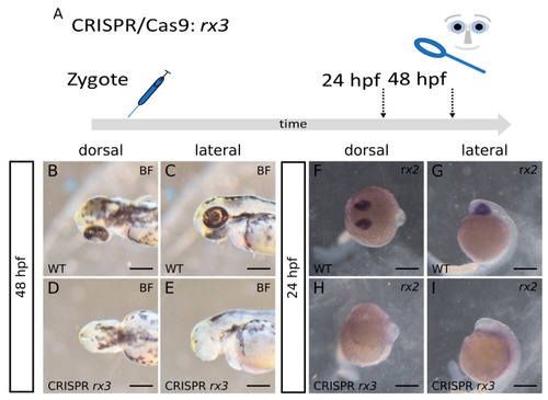
(A): summary of experimental procedure: embryos were injected at 1-cell-stage and analyzed at 24 hpf or 48 hpf. (B–E): Brightfield-images at 48 hpf. We observed anophthalmia in F0 rx3 Crispants. (F–I): ISH for rx2 at 24 hpf. The expression of rx2 is absent in F0 rx3 Crispants at 24 hpf, correlating with the severity of the phenotype. (B,C,F,G): wildtype. (D,E,H,I): rx3-crispant. (B,D,F,H) dorsal view. (C,E,G,I) lateral view. scalebars indicate 250 µm. |
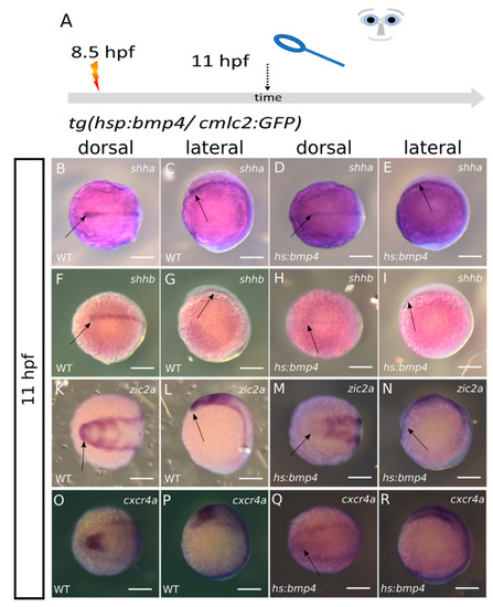
ISH at 11 hpf after bmp4-induction at 8.5 hpf. (A): summary of experimental procedure: embryos were heat-shocked at 8.5 hpf and analyzed at 11 hpf; left columns wildtypes, right columns bmp4-induced;first and third column dorsal view, second and fourth column lateral view. (B–E): Expression of shha is reduced in the area of the prechordal plate (arrows) after bmp4-induction yet still present. (F–I): shhb expression is also still present after bmp4-induction but reduced in intensity (arrows). (K–N): Expression of zic2a is absent in the ANP domain (arrows) after bmp4-induction. (O–R): expression of cxcr4a in the eye field is strongly reduced after bmp4-induction. scalebars indicate 200 µm. |
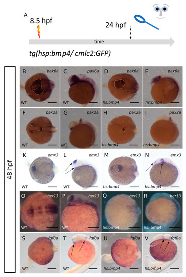
Analysis of marker genes of telencephalic, retinal and hypothalamic precursors after bmp4-induction. (A): summary of experimental procedure: embryos were heat-shocked at 8.5 hpf and analyzed at 24 hpf. Left columns wildtypes, right columns bmp4-induced embryos. First and third column dorsal view, second and fourth column lateral view. (B–E): ISH for pax6a at 24 hpf. Pax6a (dotted lines) is expressed in the crypt-oculoid and in the diencephalic domain after bmp4-induction. (F–I): ISH for pax2a (dotted lines) at 24 hpf. The expression of pax2a in the MHB is conserved after bmp4-induction but the optic stalk domain is lost. (K–N): ISH for emx3 at 24 hpf. Emx3 is present after bmp4-induction but the expression pattern is changed (arrows). (O–R): ISH for her13 at 24 hpf. The expression of her13 is lost in anterior regions after bmp4-induction. (S–V): ISH for fgf8a at 24 hpf. Two expression domains posterior to the crypt-oculoid are visible after bmp4-induction (arrows). The ventral expression domain (left arrow in T) is ceased after bmp4-induction. Scalebars indicate 200 µm. |

Scheme, summary of major findings. (A): Experimental induction of bmp4 results in a suppression of rx3 and shha/b with a consecutive suppression of cxcr4a and zic2a. The ANP is not splitting in a proper manner, resulting in a crypt-oculoid. Six3b and emx3 are found condensed at the midline. (B): scheme showing the pathological development of the ANP resulting from bmp4 induction, including a defect in the hypothalamic subduction. (C): brief summary of findings. |