
|
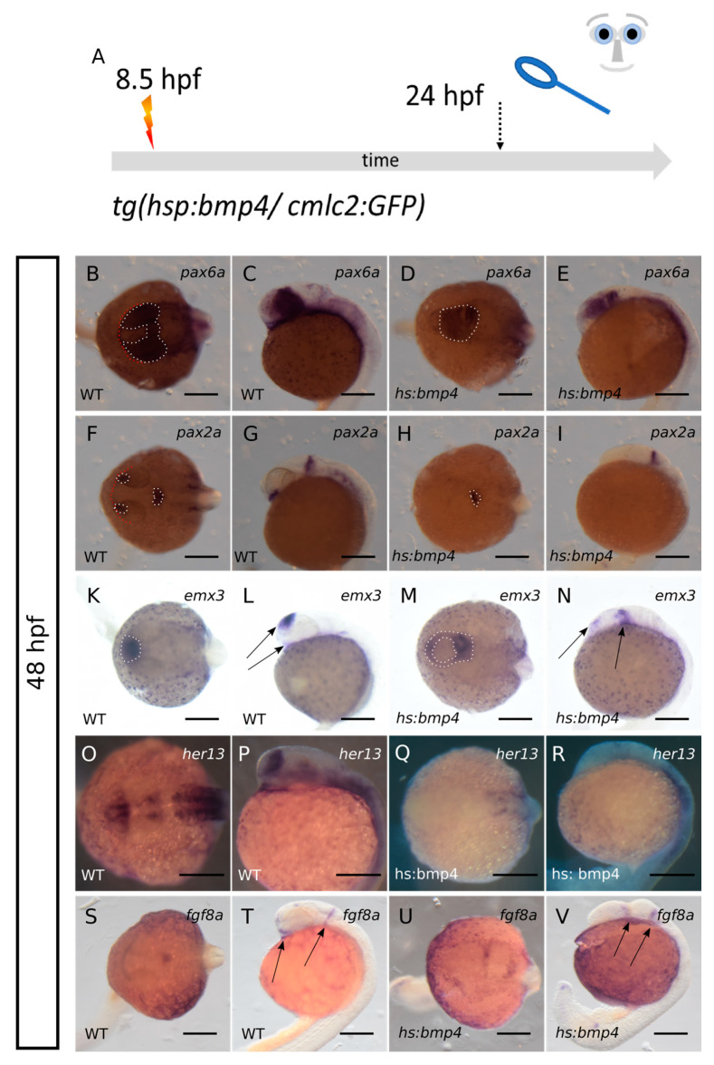
Figure 5
Analysis of marker genes of telencephalic, retinal and hypothalamic precursors after bmp4-induction. (A): summary of experimental procedure: embryos were heat-shocked at 8.5 hpf and analyzed at 24 hpf. Left columns wildtypes, right columns bmp4-induced embryos. First and third column dorsal view, second and fourth column lateral view. (B–E): ISH for pax6a at 24 hpf. Pax6a (dotted lines) is expressed in the crypt-oculoid and in the diencephalic domain after bmp4-induction. (F–I): ISH for pax2a (dotted lines) at 24 hpf. The expression of pax2a in the MHB is conserved after bmp4-induction but the optic stalk domain is lost. (K–N): ISH for emx3 at 24 hpf. Emx3 is present after bmp4-induction but the expression pattern is changed (arrows). (O–R): ISH for her13 at 24 hpf. The expression of her13 is lost in anterior regions after bmp4-induction. (S–V): ISH for fgf8a at 24 hpf. Two expression domains posterior to the crypt-oculoid are visible after bmp4-induction (arrows). The ventral expression domain (left arrow in T) is ceased after bmp4-induction. Scalebars indicate 200 µm.