
- Title
-
Zebrafish Patient-Derived Xenograft Model as a Preclinical Platform for Uveal Melanoma Drug Discovery
- Authors
- Yin, J., Zhao, G., Kalirai, H., Coupland, S.E., Jochemsen, A.G., Forn-Cuní, G., Wierenga, A.P.A., Jager, M.J., Snaar-Jagalska, B.E., Groenewoud, A.
- Source
- Full text @ Pharmaceuticals (Basel)
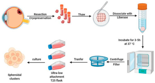
Schematic representation of patient–derived UM spheroid culture generation. The UM primary tissues were resected and frozen for storage or used directly. Tissue pieces were disaggregated, enzymatically dissociated, purified, and cultured in ultra-low adhesion plates, to form spheroids for further research. |
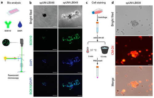
Analysis and transient chemical labelling of the patient-derived spheroids. (a) Workflow of immunostaining in the patient-derived spheroids. (b) Melanocyte marker SOX10 is expressed in spUM-LB046 and spUM-LB049. (c) Workflow of CellTracker staining of spheroids. (d) spUM-LB008 labeled using CM-Dil expressed red fluorescence and kept its spherical morphology for at least 5 days after staining. The images were taken with 20× magnification. The scale bar is 100 µm. |

Establishment of zebrafish patient-derived xenograft model. (a) The timeline of drug treatment in the zebrafish xenograft assay: collection of embryos (0 dpf), UM cell injection (2 dpf), drug administration (1–5 dpi), imaging or IHC staining (6 dpi). (b) The fluorescent images of whole zebrafish engrafted without and with spheroid-derived cells at 5 dpi. The cells disseminated into the caudal hematopoietic tissue (CHT), while some cells remained at the injection site. The white arrows in the images of spUM-LB046 and spUM-LB049 point to extravascular cells. Images were taken with 20× magnification. The scale bar is 400 µm. (c) H&E and Melan-A staining of spUM-LB046 and spUM-LB049 at 6 dpi. The Melan-A images in black magnification showed the metastasis of UM cells in the brain of engrafted zebrafish embryos. The scale bar is 500 µm. |

The maximum tolerated dose (MTD) of drug in the wild-type zebrafish. (a) Schematic diagram of the experimental set up for drug toxicity in wild-type zebrafish embryos: drug administration (3–7 dpf) and measurement of MTD (8 dpf). (b) Representative images of normal (top) and malformed (bottom) zebrafish larvae at 8 dpf. (c) The dotted line denotes the 80% survival rate used as a cut-off for the establishment of the MTD. The survival of zebrafish treated with navitoclax top exceeded 80% at concentration below 0.625 µM. The black arrow indicates the MTD of navitoclax (0.625 µM). (d) The black arrow indicates the MTD of navitoclax (0.625 µM) + everolimus (0.625 µM) when treated through bath submersion administration. |

Navitoclax and everolimus and the combination of both chemotherapeutic agents reduced spUM-LB008 tumor burden in zf-PDX model. (a) The experimental layout of drug treatment in the UM zf-PDX model. (b) Representative phenotypes of zebrafish in the DMSO-control and drug treatment groups at 6 dpi (n = 3, p < 0.01). P values were indicated as follows: ** p < 0.01, *** p < 0.001, **** p < 0.0001. (c) Effect of navitoclax, everolimus, and their combination treatment on tumor burden, compared to the control group. The combination of navitoclax and everolimus reduced the tumor burden significantly compared to everolimus and navitoclax alone. The fluorescence intensity in CHT (white rectangle) of each individual zebrafish was measured as the metastasized tumor burden. The scale bar is 500 µm. |

zf-PDX model derived from short-lived UM spheroids for drug discovery. After generation of patient-derived spheroids, ex vivo analysis was used to detect their tumorigenic and melanocytic characteristics. Transient cell staining was used to visualize the cell distribution in the zebrafish UM-PDX model. The drug screening using zf-PDX enabled evaluation of anti-UM drugs in a preclinical setting (Created with BioRender.com). |