
Image
|
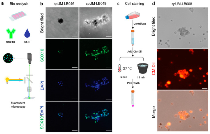
Figure Caption
Figure 2
Analysis and transient chemical labelling of the patient-derived spheroids. (a) Workflow of immunostaining in the patient-derived spheroids. (b) Melanocyte marker SOX10 is expressed in spUM-LB046 and spUM-LB049. (c) Workflow of CellTracker staining of spheroids. (d) spUM-LB008 labeled using CM-Dil expressed red fluorescence and kept its spherical morphology for at least 5 days after staining. The images were taken with 20× magnification. The scale bar is 100 µm.
Acknowledgments
This image is the copyrighted work of the attributed author or publisher, and
ZFIN has permission only to display this image to its users.
Additional permissions should be obtained from the applicable author or publisher of the image.
Full text @ Pharmaceuticals (Basel)