Flow chart for the extraction of Physalis alkekengi L. (a) and the luteolin-7,4′-di-O-β-D-glucopyranoside analyzed by HPLC (b).

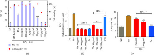
Quantification of NO production and cell viability (a), NOS activities (b), and cell apoptosis (c) in RAW264.7. PEs at different concentrations were incubated with cells for 1 h prior to incubation with LPS (1 μg/mL) for 24 h. Treatment of LPS plus Dex at 10 μg/mL was a positive control. Each experiment was conducted in triplicate. Significant differences from the LPS-treated group were regarded as  ∗∗p < 0.01,  ∗∗∗p < 0.001.

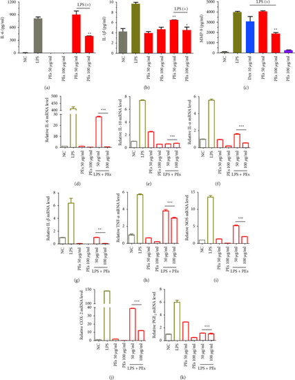
Anti-inflammatory effects of PEs on LPS-stimulated RAW264.7. Cells were pretreated with PEs at the concentrations of 50, 100 μg/mL for 1 h and exposed to 1 µg/mL LPS for another 24 h. After, IL-6 (a), IL-1β (b), and MMP-9 (c) production in the cell culture supernatant were detected by using an enzyme-linked immunosorbent assay (ELISA). The cells were collected and lysed for the measurement of the relative expression of genes involved in immune responses (IL-6 (d), IL-10 (e), IL-α (f), IL-1β (g), TNF-α (h), NOS (i), COX-2 (j), and PGE2 (k)) by qRT-PCR analysis (β-Actin expression was used as an internal control). Each experiment was performed in triplicate. p < 0.05,  ∗∗p < 0.01,  ∗∗∗p < 0.001 compared to the group of LPS-treatment alone.

Antisenescent effect of PEs on HFF-1. Cells were treated with 20 mg/mL of D-gal together with 100 ng/mL of PEs for 72 h. After, cells were stained according to the manufacturer's protocol. β-gal positive cells (in blue) were observed through microscopy (scale bar represents 20 µm) (a) and counted (b), moreover, the anti-inflammatory effect of PEs on HFF-1 was evaluated according to the ELISA experiment of IL-1β in cell culture medium after treatment with 400 µM of H2O2 with/without 100 ng/mL PEs (c). Each experiment was performed in triplicate. p < 0.01 compared to the groups of D-gal- or H2O2-treatment.

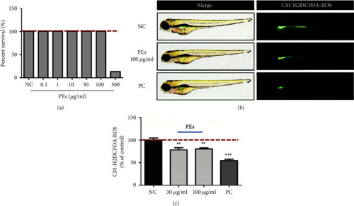
Antioxidant effect of PEs in vivo. Thirty zebrafish embryos were incubated either with PEs at different concentrations or Tripeptide-1 as the positive control (PC) for 24 h. After, toxicity in zebrafish embryos was determined through bright-field microscopy while the ROS levels were detected through a fluorescent dye H2DCFDA. Briefly, 20 μg/mL of H2DCFDA replaced the PEs to incubate with embryos for 1 h in the dark. After washing three times, the fluorescence was observed via fluorescence microscopy (c) and the fluorescence intensity was obtained using a microplate reader (b). p < 0.01, ∗∗p < 0.001 compared to the nontreatment group.

Anti-inflammatory effect of PEs against CuSO4-induced inflammation in vivo. (a) Effective concentrations of PEs and anti-inflammatory efficacies. (b) The fluorescence images of zebrafish embryos after treatment with 1 µM CuSO4 alone or together with PEs at different concentrations. (c) The number of neutrophils in transgenic zebrafish embryos under different treatment conditions, ∗∗p < 0.001 compared with the nontreatment group, and #p < 0.05, ###p < 0.001 compared with the group treated with CuSO4 alone (a-c). The expression of inflammation-related genes (IL-6, COX-2) and eicosanoid pathway-associated genes (COX-2, C3a, and PLA2) in vivo. The zebrafish embryos were exposed to CuSO4 either with PEs or not for 2h. After, embryos were collected for qRT-PCR analysis. A pool of 30 embryos per group was used. Experiments were performed in triplicates. p < 0.05, ∗∗p < 0.001 compared to the experiment treated with CuSO4 alone (d-g).

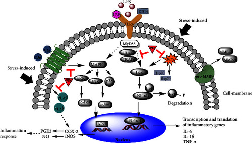
Proposed roles of PEs in the suppression of LPS-induced and oxidative stress-induced inflammatory pathways.

Acknowledgments
This image is the copyrighted work of the attributed author or publisher, and ZFIN has permission only to display this image to its users. Additional permissions should be obtained from the applicable author or publisher of the image. Full text @ Evid. Based Complement. Alternat. Med.