IMAGE

Figure 4

ID
ZDB-IMAGE-221104-5
Source
Figures for He et al., 2022
Image
Figure Caption

Figure 4

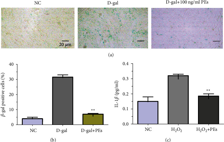
Antisenescent effect of PEs on HFF-1. Cells were treated with 20 mg/mL of D-gal together with 100 ng/mL of PEs for 72 h. After, cells were stained according to the manufacturer's protocol. β-gal positive cells (in blue) were observed through microscopy (scale bar represents 20 µm) (a) and counted (b), moreover, the anti-inflammatory effect of PEs on HFF-1 was evaluated according to the ELISA experiment of IL-1β in cell culture medium after treatment with 400 µM of H2O2 with/without 100 ng/mL PEs (c). Each experiment was performed in triplicate. p < 0.01 compared to the groups of D-gal- or H2O2-treatment.

Acknowledgments
This image is the copyrighted work of the attributed author or publisher, and ZFIN has permission only to display this image to its users. Additional permissions should be obtained from the applicable author or publisher of the image. Full text @ Evid. Based Complement. Alternat. Med.