FIGURE SUMMARY
Title

Evaluation of phages and liposomes as combination therapy to counteract Pseudomonas aeruginosa infection in wild-type and CFTR-null models

Authors
Cafora, M., Poerio, N., Forti, F., Loberto, N., Pin, D., Bassi, R., Aureli, M., Briani, F., Pistocchi, A., Fraziano, M.
Source
Full text @ Front Microbiol

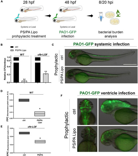
Antimicrobial activity of phosphatidylserine/phosphatidic acid (PS/PA) liposome prophylactic administration in wild-type (WT) and cftr-LOF zebrafish embryos. (A) Schematic representation of PS/PA liposome prophylactic treatment. Zebrafish embryos were treated with PS/PA liposome at 28 hpf, then systemically or locally infected with 100–300 CFU PAO1-GFP at 48 hpf and analyzed for bacterial burden at 8 or 20 h post infection. (B) Bacterial load (relative CFU/embryo) in systemically infected WT and cftr-LOF embryos, control (ctrl), and PS/PA liposome treated at 8 hpi. Results are presented as mean ± SEM. (C) Representative images of PAO1-GFP systemic bacterial infection in ctrl and PS/PA liposome treated embryos. (D,E) Quantitative analysis (fluorescence pixel count) of PAO1-GFP locally injected in the close cavity of the hindbrain ventricle of WT and cftr-LOF embryos, control (ctrl) and PS/PA liposome treated. The mean and the min to max values of at least two independent experiments (3–10 embryos/treatment) were reported on floating bars. (F) Representative images of PAO1-GFP ventricle bacterial infection in ctrl and PS/PA liposome treated embryos. Statistical significance was assessed by unpaired Student’s t test: **p < 0.01; *p < 0.05. Scale bar indicates 500 μm in panel (C) and 200 μm (dorsal) and 150 μm (lateral) in panel (F).

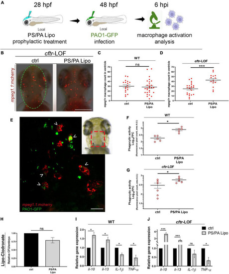
Macrophage activation in wild-type (WT) and cftr-LOF zebrafish embryos upon phosphatidylserine/phosphatidic acid (PS/PA) liposome prophylactic administration. (A) Schematic representation of PS/PA liposome prophylactic treatment. Zebrafish embryos were treated with PS/PA liposome at 28 hpf, then locally infected with PAO-GFP at 48 hpf and analyzed for macrophage activation at 6 h post infection. (B) Representative image of macrophage migration toward the PAO1-GFP bacteria injected in the hindbrain ventricle (circle area) of ctrl or PS/PA liposome treated cftr-LOF embryos. (C,D) Quantification of mpeg1:mcherry positive macrophages in the selected area of the ventricle of ctrl or PS/PA liposome treated WT (C) or cftr-LOF embryos (D). (E) Representative image of red macrophages of the Tg(mpeg1:mcherry) embryos phagocyting PAO1-GFP bacteria (arrowheads), injected in the hindbrain ventricle (visual imaging in the right-upper box). (F,G) Quantitative analysis (Log10 fluorescence pixel count, related to colocalization area) of phagocytic activity of macrophages against PAO1-GFP bacteria in WT (F) and cftr-LOF embryos (G), control (ctrl) and PS/PA liposome treated. (H,I) Pro- and anti-inflammatory cytokines expression by RT-qPCR analyses at 20 hpi in WT (I) and cftr-LOF embryos (J), ctrl and PS/PA liposome treated embryos, systemically infected with PAO1. (J) Bacterial load quantification (relative CFU/embryo) at 8 hpi in ctrl and PS/PA liposome treated WT embryos treated with Lipo-clodronate. Unpaired Student’s t test: ***p < 0.001; *p < 0.05; ns: not significant. Data resulted from at least two (C,D,F,G) or three (H–J) independent experiments and results are presented as mean ± SEM. Scale bar indicates 200 μm in panel (B) 20 μm in panel (E).

Antimicrobial activity in WT and cftr-LOF zebrafish embryos of the combination treatment with phosphatidylserine/phosphatidic acid (PS/PA) liposome in prophylactic administration and phage therapy. (A) Schematic representation of PS/PA liposome prophylactic treatment followed by systemic bacterial infection and CKΦ-administration. Zebrafish embryos were treated with PS/PA liposome at 28 hpf, then systemically infected with PAO1-GFP at 48 hpf, treated with CKΦ 3 h later, and analyzed for bacterial burden at 8 or 20 h post infection. (B,C) Bacterial load (relative CFU/embryo) in systemically infected wild-type (WT) (B), and cftr-LOF embryos (C), control (ctrl), PS/PA liposome, CKΦ and PS/PA liposome + CKΦ treated. Statistical significance was assessed by One-way ANOVA followed by Tukey’s post hoc test: ***p < 0.001; *p < 0.05; ns, not significant. Data resulted from three independent experiments and results are presented as mean ± SEM.

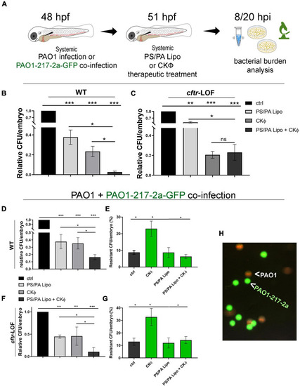
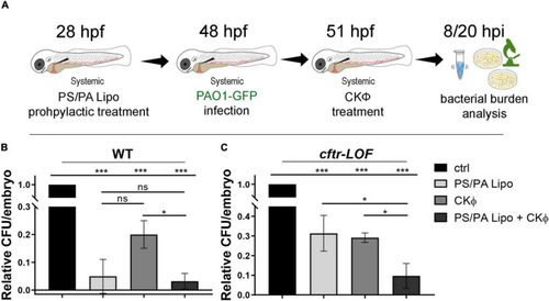
Combination treatment with phosphatidylserine/phosphatidic acid (PS/PA) liposome and CKΦ in therapeutic administration elicits synergistic effect in antimicrobial activity in wild-type (WT) and decreases phage-resistant PAO1 proliferation in cftr-LOF zebrafish embryos. (A) Schematic representation of combined administration of PS/PA liposome/CKΦ. 48 hpf zebrafish embryos were systemically infected with phage sensitive (PAO1) and/or resistant PAO1 (PAO1-217-2a-GFP) strains, treated with single or combined PS/PA liposome CKΦ 3 h later and analyzed for bacterial burden at 8 or 20 h post infection. (B,C) Bacterial load (relative CFU/embryo) in systemically infected WT (B) and cftr-LOF embryos (C), control (ctrl), PS/PA liposome, CKΦ and PS/PA liposome-CKΦ treated at 8 hpi. (D–G) Bacterial load (relative CFU/embryo) (D,F) and percentage of PAO1-217-2a-GFP colonies (E,G) in embryos systemically infected with 50% phage-sensitive PAO1 (non-GFP) and 50% phage-resistant PAO1-GFP bacterial suspension. WT (D,E) or in cftr-LOF embryos (F,G), control (ctrl), PS/PA liposome, CKΦ and PS/PA liposome-CKΦ treated. (H) Representative image of phage-sensitive PAO1 and phage resistant PAO1-217-2a-GFP colonies derived from the plating of homogenized infected embryos. Statistical significance was assessed by One-way ANOVA test followed by Tukey’s post hoc correction:***p < 0.001; **p < 0.01; *p < 0.05; ns, not significant. Data resulted from three independent experiments and results are presented as mean ± SEM.

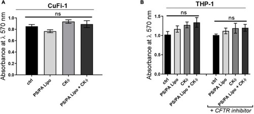
PS/PA liposomes and CKΦ administration does not elicit toxic effects on human CF cells. (A) Cell viability, evaluated by MTT assay, in immortalized human bronchial epithelial cells CuFi-1 expressing F508del CFTR control, treated with phosphatidylserine/phosphatidic acid (PS/PA) liposome, CKΦ or combination of PS/PA liposome-CKΦ for 48 h. (B) Cell viability, evaluated by MTT assay, in THP-1 activated macrophage-like cells treated or not with the CFTR inhibitor. Effects of single or combined PS/PA liposome and CKΦ-administration were evaluated after 48 h of treatment. Unpaired Student’s t test: *p < 0.05; ns, not significant. Results are presented as mean ± SEM.

Acknowledgments
This image is the copyrighted work of the attributed author or publisher, and ZFIN has permission only to display this image to its users. Additional permissions should be obtained from the applicable author or publisher of the image. Full text @ Front Microbiol