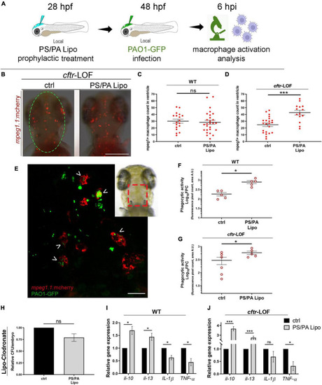
Macrophage activation in wild-type (WT) and cftr-LOF zebrafish embryos upon phosphatidylserine/phosphatidic acid (PS/PA) liposome prophylactic administration. (A) Schematic representation of PS/PA liposome prophylactic treatment. Zebrafish embryos were treated with PS/PA liposome at 28 hpf, then locally infected with PAO-GFP at 48 hpf and analyzed for macrophage activation at 6 h post infection. (B) Representative image of macrophage migration toward the PAO1-GFP bacteria injected in the hindbrain ventricle (circle area) of ctrl or PS/PA liposome treated cftr-LOF embryos. (C,D) Quantification of mpeg1:mcherry positive macrophages in the selected area of the ventricle of ctrl or PS/PA liposome treated WT (C) or cftr-LOF embryos (D). (E) Representative image of red macrophages of the Tg(mpeg1:mcherry) embryos phagocyting PAO1-GFP bacteria (arrowheads), injected in the hindbrain ventricle (visual imaging in the right-upper box). (F,G) Quantitative analysis (Log10 fluorescence pixel count, related to colocalization area) of phagocytic activity of macrophages against PAO1-GFP bacteria in WT (F) and cftr-LOF embryos (G), control (ctrl) and PS/PA liposome treated. (H,I) Pro- and anti-inflammatory cytokines expression by RT-qPCR analyses at 20 hpi in WT (I) and cftr-LOF embryos (J), ctrl and PS/PA liposome treated embryos, systemically infected with PAO1. (J) Bacterial load quantification (relative CFU/embryo) at 8 hpi in ctrl and PS/PA liposome treated WT embryos treated with Lipo-clodronate. Unpaired Student’s t test: ***p < 0.001; *p < 0.05; ns: not significant. Data resulted from at least two (C,D,F,G) or three (H–J) independent experiments and results are presented as mean ± SEM. Scale bar indicates 200 μm in panel (B) 20 μm in panel (E).
|
