IMAGE

FIGURE 1

ID
ZDB-IMAGE-221004-33
Source
Figures for Cafora et al., 2022
Image
Figure Caption

FIGURE 1

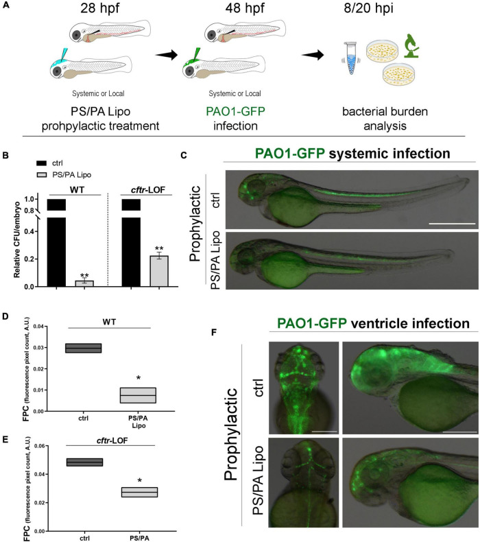
Antimicrobial activity of phosphatidylserine/phosphatidic acid (PS/PA) liposome prophylactic administration in wild-type (WT) and cftr-LOF zebrafish embryos. (A) Schematic representation of PS/PA liposome prophylactic treatment. Zebrafish embryos were treated with PS/PA liposome at 28 hpf, then systemically or locally infected with 100–300 CFU PAO1-GFP at 48 hpf and analyzed for bacterial burden at 8 or 20 h post infection. (B) Bacterial load (relative CFU/embryo) in systemically infected WT and cftr-LOF embryos, control (ctrl), and PS/PA liposome treated at 8 hpi. Results are presented as mean ± SEM. (C) Representative images of PAO1-GFP systemic bacterial infection in ctrl and PS/PA liposome treated embryos. (D,E) Quantitative analysis (fluorescence pixel count) of PAO1-GFP locally injected in the close cavity of the hindbrain ventricle of WT and cftr-LOF embryos, control (ctrl) and PS/PA liposome treated. The mean and the min to max values of at least two independent experiments (3–10 embryos/treatment) were reported on floating bars. (F) Representative images of PAO1-GFP ventricle bacterial infection in ctrl and PS/PA liposome treated embryos. Statistical significance was assessed by unpaired Student’s t test: **p < 0.01; *p < 0.05. Scale bar indicates 500 μm in panel (C) and 200 μm (dorsal) and 150 μm (lateral) in panel (F).

Acknowledgments
This image is the copyrighted work of the attributed author or publisher, and ZFIN has permission only to display this image to its users. Additional permissions should be obtained from the applicable author or publisher of the image. Full text @ Front Microbiol