- Title
-
Ifi30 Is Required for Sprouting Angiogenesis During Caudal Vein Plexus Formation in Zebrafish
- Authors
- Wang, X., Ge, X., Qin, Y., Liu, D., Chen, C.
- Source
- Full text @ Front. Physiol.
|
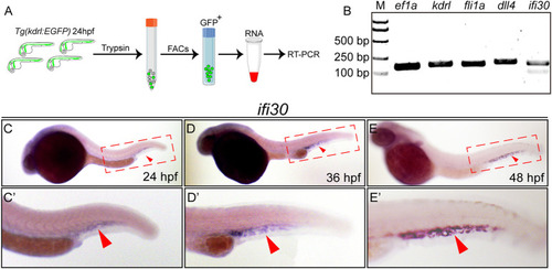
ifi30 enriches in endothelial cells (ECs) and expresses in caudal vein plexus (CVP) region (A) Schematic representation of isolation of ECs from Tg(kdrl:EGFP) zebrafish embryos for RNA extraction and RT-PCR (B) Expressions of kdrl, fli1a, dll4, and ifi30 in ECs are examined by RT-PCR. ef1a is used as the housekeeping gene. M indicates the DNA ladder and the corresponding sizes are marked on the left (C–E) Expression of ifi30 in zebrafish embryos from 24 to 48 hpf (C′-E′) Magnifications of the red dotted boxes in the corresponding (C) (D), and (E) panel, respectively. Ifi30 is specially expressed in the CVP region (indicated with red arrowhead). |
|
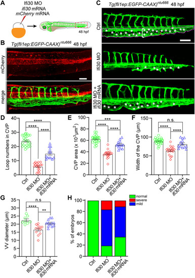
Ifi30 overexpression rescues the defective CVP phenotypes of Ifi30 morphants (A) Schematic representation of the rescue experiment. The mixture of Ifi30 MO, ifi30 mRNA, and mCherry mRNA is injected into one-cell stage Tg(fli1ep:EGFP-CAAX) ntu666 embryos. The mCherry mRNA is used to verify the working efficiency of mRNA injection by examining the red fluorescence post injection (B) (C–G) The CVP loop numbers (asterisks), total CVP area (white dotted line outlined area), CVP width (white square bracket), and ventral vein (VV) diameter (red square bracket) in Ifi30 morphants are all rescued by ifi30 mRNA injection (H) The incidence of normal and defective CVP phenotypes in control Tg(fli1ep:EGFP-CAAX) ntu666 embryos (n = 34), embryos injected with Ifi30 MO (n = 41), and embryos co-injected with Ifi30 MO and ifi30 mRNA (n = 34). Error bars represent SEM. n. s., not significant. **, p < 0.01. ***, p < 0.001. ****, p < 0.0001. Scale bars, 100 µm. |
|
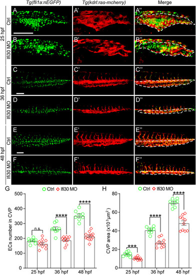
Ifi30 loss-of-function impairs endothelial cell (EC) proliferation during CVP development (A–F") Confocal images of Tg(kdrl:ras-mCherry);Tg(fli1:nEGFP) double transgenic embryos and embryos injected with Ifi30 MO at 25 hpf (A,B"), 36 hpf (C,D"), and 48 hpf (E,F"). The EC numbers in the CVP region and CVP area (white dotted line outlined area) were quantified (G,H). Error bars represent SEM. n. s., not significant. ***, p < 0.001. ****, p < 0.0001. Scale bars, 100 µm. |
|
Ifi30 knockdown disrupts Bmp2 signaling pathway. In situ hybridization for bmp2b, bmpr2a, and dab2 of control embryos and embryos injected with Ifi30 MO at 24 and 36 hpf. |
|
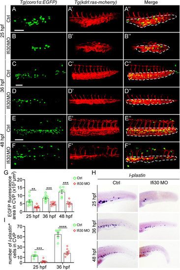
Macrophages are reduced upon Ifi30 knockdown (A–F") Confocal images of Tg(kdrl:ras-mCherry);Tg(coro1a:EGFP) double transgenic embryos and embryos injected with Ifi30 MO at 25 hpf (A,B"), 36 hpf (C,D"), and 48 hpf (E,F") (G) Fluorescence intensities in the CVP region (dashed line outlined area) of GFP channel are quantified with ImageJ (H) In situ hybridization (ISH) assay of l-plastin (macrophage marker) in wild-type (WT) and Ifi30 morphant embryos at 25, 36, and 48 hpf, respectively. The number of l-plastin-positive cells in the CVP region (dashed line outlined area) is counted and compared between WT and Ifi30 morphants (I). Error bars represent SEM. **, p < 0.01. ***, p < 0.001. ****, p < 0.0001. Scale bars, 100 µm. |

