
IMAGE
Fig. 4
- ID
- ZDB-IMAGE-220802-38
- Publication
- Wang et al., 2022 - Ifi30 Is Required for Sprouting Angiogenesis During Caudal Vein Plexus Formation in Zebrafish
- All Figures
- Figures for Wang et al., 2022
Image
|
Figure Caption
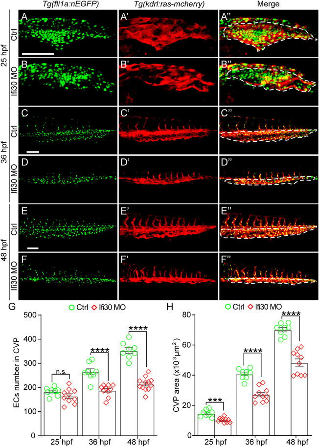
Fig. 4
Ifi30 loss-of-function impairs endothelial cell (EC) proliferation during CVP development (A–F") Confocal images of Tg(kdrl:ras-mCherry);Tg(fli1:nEGFP) double transgenic embryos and embryos injected with Ifi30 MO at 25 hpf (A,B"), 36 hpf (C,D"), and 48 hpf (E,F"). The EC numbers in the CVP region and CVP area (white dotted line outlined area) were quantified (G,H). Error bars represent SEM. n. s., not significant. ***, p < 0.001. ****, p < 0.0001. Scale bars, 100 µm.
Acknowledgments
This image is the copyrighted work of the attributed author or publisher, and
ZFIN has permission only to display this image to its users.
Additional permissions should be obtained from the applicable author or publisher of the image.
Full text @ Front. Physiol.