FIGURE SUMMARY
Title

The Olfactory Organ Is a Unique Site for Neutrophils in the Brain

Authors
Palominos, M.F., CalfĂșn, C., Nardocci, G., Candia, D., Torres-Paz, J., Whitlock, K.E.
Source
Full text @ Front Immunol

Neutrophils are found only in the olfactory organs of the adult brain. (A) Wholemount brain of Tg(omp:RFP);Tg(mpx:GFP) adult: neutrophils (green) are only present in the OO (OE/EN); Tel: telencephalon; pi: pineal. Scale bars A, B = 200 μm. (B) OO (from A) contains a large population of neutrophils (green, n=487 neutrophils). omp:RFP+ OSNs are located only in sensory epithelia (ss, red) not in non-sensory epithelia (ns). ON: olfactory nerve. (C) Neutrophils with a rounded shape, (nt1, arrow: circularity index 0.7 or greater) and amoeboid shape (nt2, arrow: circularity index of 0.4-0.6; F) were observed in the LOE. (D) Neutrophils with an amoeboid shape (nt2, arrow) were located throughout the OE and EN. (D1) Sustentacular-like cells (D1), circularity index 0.2, lie at ns-ss epithelia interface (see Figure 2). (E) Total number of neutrophils in the OO. The non-sensory (ns) tissues (respiratory epithelia + NE, blue) have more neutrophils than sensory epithelia (ss, red), n= 3 OE from 3 different fish. (E) **indicates significant statistical differences between means with a p-value < 0.002, paired t-test.. (F) Frequency distribution of nt1 and nt2 cells (n= 53 neutrophils from brain shown in C). (G) Maximal projection of whole mount Tg(mpx:GFP) adult OE: Neutrophils (green); autofluorescence (gray). (H) Neutrophils (from G) were color-coded based on (H) Z-stack depth. Total depth= 550μm. Scale bars A, B = 200 μm; C = 60 μm; D, D1 = 20 μm; G, H =100 μm. (A, B) 9 brains imaged; (C, D) 6 brains sectioned.

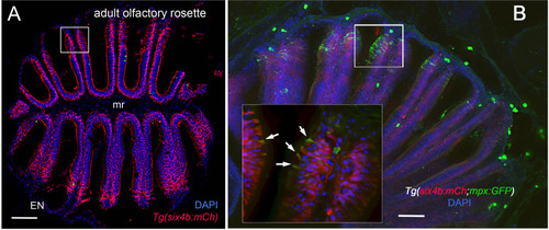
Sustentacular cells in the OE are associated with markers for neutrophils. (A, B) Cryosections of adult olfactory epithelia. (A) Adult olfactory rosette (OE) from Tg(six4b:mCh) line showing sustentacular cells (red) that are distributed within the lamellae of the OE where some areas have denser clusters (boxed area). EN: Epineurium; mr: midline raphe. Scale bar = 100 μm. (B) Sectioned OE of Tg(six4b:mCh;mpx:GFP) Six4b:mCh+ SCs (red) and mpx:GFP+ neutrophils(green) are found in the respiratory OE near the tips of the lamellae (boxed area). Scale bar = 50 μm. Inset B:. A small subset on cells are positive are both mpx:GFP+ and Six4b:mCh+ positive (arrows). (A) 1 sectioned brain, (B) 2 sectioned brains.

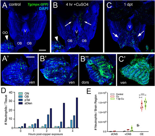
Exposure to copper is correlated with increased neutrophils in the peripheral and central nervous system. (A–C) Ventral views of whole mount adult brains from Tg(mpx:GFP). Scale bars: A-C; A’-C’= 100 μm. (A, A’) Control with neutrophils found only in OO (arrowhead; A’). (B, B’, B”) After four-hour exposure to copper, there was an increase in the number of neutrophils in the OO (B, arrowhead, B’, B”). In addition, neutrophils were observed in the ventral OB, along the ventricle (V) and in the ventral telencephalon (B, arrows) (C, C’) One day post treatment neutrophils were still present in OO (C’), the OB (arrows) and the ventral telencephalon. (D) Neutrophils appear over time in an anterior to posterior spatial pattern: olfactory nerve (ON), olfactory bulb (OB), posterior Telencephalon (pTel), and anterior diencephalon; (the numbers for OO are not plotted because number is out of range (average ~1,500, see E). (E) Copper exposure was correlated with increased neutrophils in OE and ventral CNS. ****: Two-way ANOVA, Tukey’s multiple comparison test, p < 0.05). (****P, 0.0001). (A–C, A’–C’) Preparations were selected for imaging based on whether they were intact and the signal to noise of the labeling. (D) 1 brain was analyzed per timepoint. (E) 3 brains were examined per treatment.

Damage induces cell proliferation of OSN and of neutrophil precursors in the adult olfactory organ. (A–C) BrdU labeled cells (white), neutrophils (green) in whole mount OO of adult fish. Scale bars A-C = 50 μm (A). Prior to copper exposure BrdU labeling and scattered neutrophils were observed in the medial raphe (mr), sensory (ss), and non-sensory (ns) epithelia. n= 3 OE. (B) After four hours of exposure to copper, intense BrdU labeling was observed in the mr. n= 3 OE. (C) One day post recovery, neutrophils lined the lamellae and intense BrdU labeling was observed in ss and LOE-EN. n= 3 OE. (D) Section of Tg(fli1a:EGFP) adult OE showing extensions of blood vasculature (green) within the OE. Scale bar = 100 μm. n= 3 sectioned heads. (E) Significant increases in neutrophil number were observed after 4 hour copper exposure (red) in both the ns and ss epithelia when compared to control (grey). At one dpt (green) only the number of labeled ss cells remained significantly greater than controls. n= 3 OE. (F) Damage induced changes in the number of BrdU-positive cells were significant in the ss epithelia but not the ns at 4 hour copper exposure (red) and one dpt (green). n= 3 OE. (G) A small but significant increase in mpx:GFP+ cells double labeled for BrdU scored in the OE. (E-G, n=3 adult OE from different fish; Two-way ANOVA, Tukey multiple comparison test, p < 0.05). (*P, 0.05, **P, 0.01, ***P, 0.001). n= 3 OE. (H) Four hours of copper exposure (orange) and one day post-treatment (green), an increase in rounded (nt1; circularity index 0.7 or greater see Figure 5) and amoeboid (nt2; circularity index 0.4-0.6) neutrophils were observed. Two-way ANOVA, Tukey’s multiple comparison test, p < 0.05). (*P, 0.05, **P, 0.01, ***P, 0.001, ****P, 0.0001, ns: non significant).

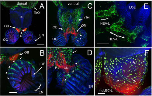
The olfactory organs have an extensive lymphatic vasculature. (A-F) Whole mount brains of adult lyve1b:EGFP;omp:RFP animals showing OSN (red) and lymphatic vasculature (green). (A) The OE and OBs but not dorsal the telencephalon (Tel) showed extensive lymphatic vasculature (LV, green). (B) Higher magnification of OO in A with the epineurium (EN, arrow) seen wrapping around the outer surface of lamella of the OE (LOE). Lymphatic cells were found in OO (arrowheads) and OB. (C) The LV extended centrally from the OO/OB along the ventral telencephalon (vTel) and posteriorly to the ventral diencephalon. (D) Higher magnification of OO in (B). LV (arrowheads) is associated with the olfactory nerve (ON, red) and covers the ventral surface of the OB (green, arrows). (E) Lyve1b:EGF+ cells in the tips of the LOE resemble High Endothelial Venules (HEVs). (F) Putative Mural lymphatic endothelial cells (MuLECs) wrap the OB (arrows). Representative images selected from detailed analysis of 9 brains. DAPI (blue). (A, C)= 200 μm; (B, D) = 100 μm; (C, F) = 50 μm.

Blood (BV) and Lymphatic (LV) Vasculature wrap the olfactory organs (OO). (A, B) Whole mount Tg(fli1a:EGFP;lyve1b:DsRed) of the adult OO connected to OB with BV (fli1a:EGFP, green) and LV (lyve1b:DsRed, red). Dorsal (A) and ventral (B) views; DAPI (blue), Scale Bars: (A, B) = 200 μm. (C) BV (red) and LV (green) density is greater in olfactory system (OS = OE and OB) than telencephalon (Tel); (SE, P-value <0.05, unpaired t-test; n = 6 adult brains. One-way ANOVA, Tukey multiple comparison test, P < 0.05). Representative images selected from detailed analysis of least 6 brains. (D) Transmitted light image of fixed whole mount OO. Boxed area corresponds to area where LOE connects with EN. Scale bar = 100 μm, (E). The LV (red) meet the BV (green) at the distal tips of each lamellae (boxed area; asterisks). Scale bar = 100 μm, (F). Cells express both lyve1b:DsRed and fli1a:EGFP (arrows). Scale bar = 25 μm. (A, B), (E, F): Analysis of 9 brains. Two-way ANOVA, Tukey’s multiple comparison test, p < 0.05). (*P, 0.05, ***P, 0.001, ns: non significant).

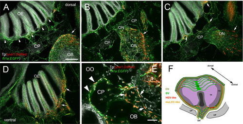
Blood vasculature extends through cribriform plate with muLEC-like lymphatic cells. (A–E) Sections from Tg(lyve1b:DsRed;fli1a:EGFP) adult brains (n=6 brains examined). (A) I. n dorsal sections the OB is separated from the OE by the cribriform plate (CP). The OB has extensive BV (green) extending into the lamellae of the OE and muLEC-like cells (red) on the surface of the OB (arrow). (A–D) = 100 μm (B). The ON passes through the CP accompanied by extensive BV (green). muLEC-like cells are on the medial surface (red, arrow) of the OB. (C). The muLEC-like cells (red, arrow) line the ventral side of the ON. (D) muLEC-like cells line the basal OE (red, arrows) in the most ventral region of the OO. (E) muLEC-like cells on the BV extending across the CP and many are positive for both lyvel1:DsRed and fli1a:EGFP (arrows). DAPI: white. Scale bar = 50 μm. (F) Diagram depicting olfactory organ with sensory (ss) and non-sensory (ns) epithelia that have extensive BV (green). The lamellae of the OE contain HEV-like LV (red) that do not extend across the cribriform plate (CP). muLEC-like cells (orange) line the BV and extend from the olfactory bulb across the CP to the basal OE. Scale bars: A-D = 100 μm, E = 50 μm.

Olfactory organs have unique neutrophil population. Schematic of olfactory organs (OOs), olfactory bulbs (OB) and telencephalon highlighting the association of the neutrophils (green) with the olfactory sensory neurons (purple). Neutrophils PC in the OO appeared in the CNS associated with posterior OB and ventricle.

Acknowledgments
This image is the copyrighted work of the attributed author or publisher, and ZFIN has permission only to display this image to its users. Additional permissions should be obtained from the applicable author or publisher of the image. Full text @ Front Immunol