IMAGE

Figure 5

ID
ZDB-IMAGE-220616-5
Source
Figures for Palominos et al., 2022
Image
Figure Caption

Figure 5

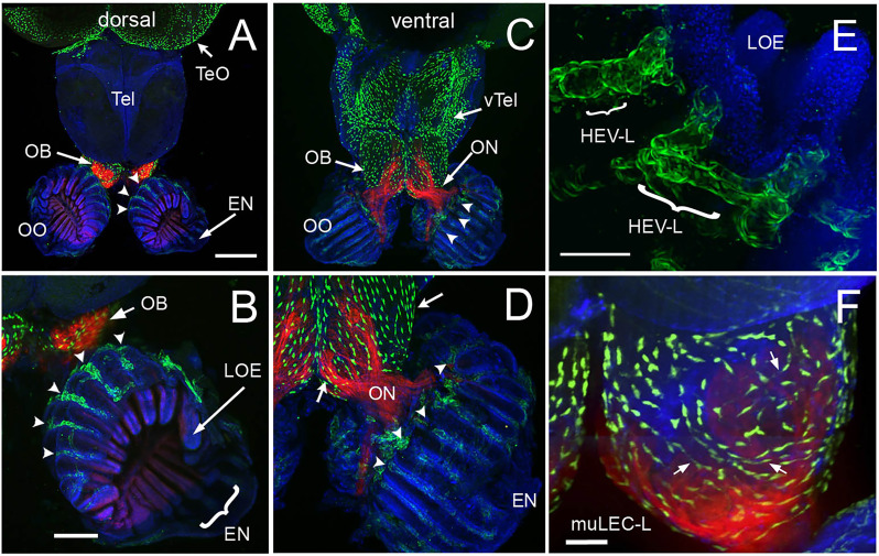
The olfactory organs have an extensive lymphatic vasculature. (A-F) Whole mount brains of adult lyve1b:EGFP;omp:RFP animals showing OSN (red) and lymphatic vasculature (green). (A) The OE and OBs but not dorsal the telencephalon (Tel) showed extensive lymphatic vasculature (LV, green). (B) Higher magnification of OO in A with the epineurium (EN, arrow) seen wrapping around the outer surface of lamella of the OE (LOE). Lymphatic cells were found in OO (arrowheads) and OB. (C) The LV extended centrally from the OO/OB along the ventral telencephalon (vTel) and posteriorly to the ventral diencephalon. (D) Higher magnification of OO in (B). LV (arrowheads) is associated with the olfactory nerve (ON, red) and covers the ventral surface of the OB (green, arrows). (E) Lyve1b:EGF+ cells in the tips of the LOE resemble High Endothelial Venules (HEVs). (F) Putative Mural lymphatic endothelial cells (MuLECs) wrap the OB (arrows). Representative images selected from detailed analysis of 9 brains. DAPI (blue). (A, C)= 200 μm; (B, D) = 100 μm; (C, F) = 50 μm.

Acknowledgments
This image is the copyrighted work of the attributed author or publisher, and ZFIN has permission only to display this image to its users. Additional permissions should be obtained from the applicable author or publisher of the image. Full text @ Front Immunol