FIGURE SUMMARY
Title

The distal C terminus of the dihydropyridine receptor β1a subunit is essential for tetrad formation in skeletal muscle

Authors
Dayal, A., Perni, S., Franzini-Armstrong, C., Beam, K.G., Grabner, M.
Source
Full text @ Proc. Natl. Acad. Sci. USA

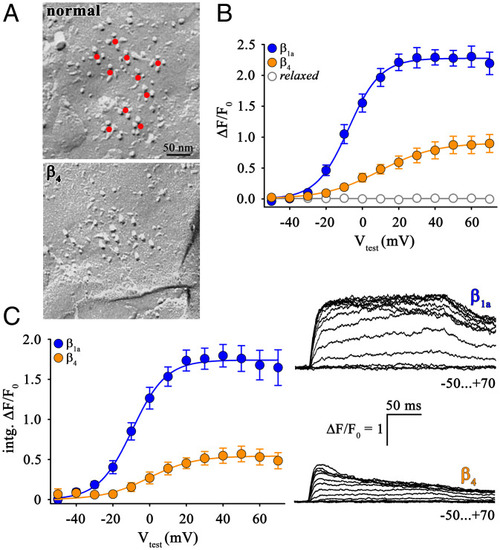
Absence of DHPR tetrad restoration in β4-expressing relaxed myotubes. (A) Freeze-fracture replicas of peripheral couplings in tail myotomes of 27- to 30-hpf zebrafish. Control myotomes (Top) show arrangement of DHPR particles in tetrads (center indicated by red dots), organized in orthogonal arrays. In β4-expressing relaxed zebrafish (Bottom) DHPR tetrads show a lack of tetrad formation. (Scale bar, 50 nm.) (B) Quantification of voltage dependence of cytoplasmic Ca2+ transients yielded (ΔF/F0)max values that are significantly lower (P < 0.001) in β4 (n = 13)- compared to β1a (n = 9)-expressing relaxed myotubes. ΔF/F0 values recorded from untransfected relaxed myotubes were below detection level (n = 10). (C) Similarly, plots of voltage dependence of the integral of the ΔF/F0 transients in response to 200-ms test depolarizations indicate a highly significant difference (P < 0.001) in the total amount of Ca2+ released between relaxed myotubes expressing β1a (n = 9) or β4 (n = 12) subunit. (Right) Representative ΔF/F0 recordings from relaxed myotubes expressing β1a or β4. (Scale bars, 50 ms [horizontal], ΔF/F0 = 1 [vertical].) Error bars indicate SEM. P determined by unpaired Student’s t test.

Loss-of-function β1a4 chimeras revealed the importance of the β1a C terminus in skeletal muscle DHPR–RyR1 coupling. (A) Block schemes of domain organization of putative loss-of-function β1a4 chimeras with systematic exchange of N terminus (N), SH3 domain (SH3), HOOK region (H), GK domain (GK), or C terminus (C) of β1a (blue) by β4 sequences (orange). Homologous SH3 and GK domains are represented by hatched boxes. (B, Left) Analyses of voltage dependence of integrated outward gating currents normalized to cell capacitance exhibited maximum charge movement (Qmax) values indistinguishable (P > 0.05) between relaxed myotubes expressing β1a (n = 16), β4 (n = 12), β1a4(N) (n = 21), β1a4(SH3) (n = 19), β1a4(H) (n = 24), β1a4(GK) (n = 15), or β1a4(C) (n = 13). Qmax values from untransfected relaxed myotubes were slightly above detection level (P < 0.001, n = 11). (Right) Representative Q recordings from relaxed myotubes expressing either β1a or β4. (Scale bars, 5 ms [horizontal], 3 pA/pF [vertical].) (C and D) Cytoplasmic Ca2+ transient restoration was comparable (P > 0.05) between relaxed myotubes expressing β1a (n = 9), β1a4(SH3) (n = 17), β1a4(GK) (n = 11), β1a4(N) (n = 14), or β1a4(H) (n = 16). By contrast, ΔF/F0 values were significantly lower (P < 0.001) for chimera β1a4(C) (n = 12) and similar (P > 0.05) to those of β4 (n = 13). Exemplar Ca2+ transient recordings from relaxed myotubes expressing β1a4(SH3) (C, Right) or β1a4(C) (D, Right). (Scale bars, 50 ms [horizontal], ΔF/F0 = 1 [vertical].) Error bars indicate SEM. P determined by unpaired Student’s t test, ***P < 0.001.

Length of the β4 C terminus is not crucial for skeletal muscle DHPR–RyR1coupling. (A) Amino acid sequence alignment depicting variable lengths of the C termini of β1a and β4 subunits (GenBank accession nos.: rabbit β1a, M25514; rat β4, L02315). To determine whether the length of the C terminus was functionally critical, the last 51 amino acids from the β4 C terminus were deleted, yielding mutant β4(Δ51). (B) Qmax values were indistinguishable (P > 0.05) between relaxed myotubes expressing the deletion mutant β4(Δ51) (n = 17), β1a (n = 16), or β4 (n = 12). (C) Maximal Ca2+ transients (ΔF/F0)max for β4(Δ51) expressing relaxed myotubes (n = 10) was similar (P > 0.05) to that of β4 (n = 13). (D) Similarly, total maximal Ca2+ transients (intg.(ΔF/F0)max) for β4(Δ51) expressing relaxed myotubes was statistically indistinguishable (P > 0.05) from that of β4. Error bars indicate SEM. P determined by unpaired Student’s t test.

The distal C terminus of β1a is crucial for skeletal muscle EC coupling. (A) Block scheme of domain organization of gain-of-function chimera β41a(C), where the the C terminus of β4 (orange) was replaced by a corresponding β1a sequence (blue). (B) Qmax values in relaxed myotubes expressing either chimera β41a(C) (n = 18) or β1a (n = 16) were comparable (P > 0.05). (C) Quantification of voltage dependence of cytoplasmic Ca2+ transients yielded significantly higher (P < 0.001) (ΔF/F0)max values for chimera β41a(C) (n = 12) compared to β4 (n = 13) but indistinguishable (P > 0.05) from that of β1a (n = 9) expressing relaxed myotubes. (Right) Exemplar cytoplasmic Ca2+ transient recordings from relaxed myotubes expressing chimera β41a(C). (Scale bars, 50 ms [horizontal], ΔF/F0 = 1 [vertical].) (D) Amino acid sequence alignment of C termini of β1a and β4 depicting the homologous proximal C terminus (green box) and heterologous distal C terminus (blue box). Red bracket indicates the highly homologous sequence in the distal C terminus of β1a revealed from sequence alignments of β1a from several vertebrate species (fish to mammals) (SI Appendix, Fig. S2B). (E) Block scheme of domain organization of chimeras β41a(prox.C) and β41a(dist.C), where the proximal and distal C terminus of β4 (orange) were exchanged by corresponding β1a sequences (blue). (F) Qmax values were indistinguishable (P > 0.05) between relaxed myotubes expressing chimera β41a(prox.C) (n = 11), β41a(dist.C) (n = 19), or β1a (n = 16). (G) Quantification of voltage dependence of cytoplasmic Ca2+ transients yielded (ΔF/F0)max values that were significantly lower (P < 0.001) for chimera β41a(prox.C) (n = 15)- compared to β1a (n = 9)-expressing relaxed myotubes. However, relaxed myotubes expressing chimera β41a(dist.C) (n = 14) exhibited pronounced Ca2+ transients, equivalent (P > 0.05) to β1a transfected myotubes (n = 13). (Right) Exemplar Ca2+ transient recordings from relaxed myotubes expressing chimera β41a(dist.C) or β41a(prox.C). (Scale bars, 50 ms [horizontal], ΔF/F0 = 1 [vertical].) (H) Quantification of spontaneous or touch-evoked coiling of 27- to 30-hpf relaxed zebrafish injected with β1a (n = 35), β4 (n = 202), β41a(C) (n = 79), and β41a(dist.C) (n = 58) mRNA. Degree of motility was indistinguishable (P > 0.05) between relaxed zebrafish expressing β41a(C) or β1a. Relaxed zebrafish expressing β41a(dist.C) displayed robust spontaneous coiling only slightly lower (P = 0.02) than β1a. Conversely, β4-injected relaxed zebrafish showed either no (n = 151) or very weak (n = 51) coiling following tactile stimulation and thus, highly significantly lower motility compared to (P < 0.001) β1a-expressing relaxed zebrafish. Uninjected relaxed zebrafish displayed neither spontaneous nor tactile-induced motility (P < 0.001, n = 28). Error bars indicate SEM. P determined by unpaired Student’s t test, *P < 0.05; ***P < 0.001.

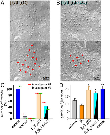
The distal C terminus of β1a is crucial for DHPR tetrad formation. (A and B) Representative freeze-fracture replicas from tail muscle tissue of 27- to 30-hpf relaxed zebrafish expressing β41a(C) (A) or β41a(dist.C) (B) reveal accurate arrangement of DHPR particles in tetrads. The red dots (Bottom) indicate the centers of three- or four-particle tetrads and additional particles that are in the expected position for an orthogonal array. (Scale bar, 50 nm.) (C) Numbers of tetrads (three or four particles) determined by two independent investigators from 95 anonymized freeze-fracture images acquired from zebrafish tails, either normal controls (normal), uninjected (relaxed), or injected with β4, β41a(C), or β41a(dist.C) mRNA. Each bar represents mean of the counts normalized to normal zebrafish (where the mean of the two investigators’ counts was defined as 100%) and the two arrows (red and green) depict the counts of the two individual investigators (SI Appendix, Table S2). (D) Counts of DHPR particles per junction from zebrafish tails, either uninjected (relaxed), injected with β4, β41a(C), or β41a(dist.C) mRNA, or normal controls (normal). Error bars indicate SEM. P determined by unpaired Student’s t test, *P < 0.05; **P < 0.01.

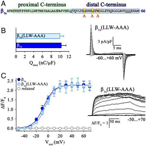
Hydrophobic residues (L496L500W503) in the β1a distal C terminus are not important for skeletal muscle EC coupling. (A) Amino acid sequence of rabbit β1a C terminus depicting the position of the three hydrophobic residues LLW (red box with yellow filling), which were exchanged with alanines (AAA). (B) Relaxed myotubes expressing triple mutant β1a(LLW-AAA) (n = 16) displayed Qmax values similar (P > 0.05) to β1a (n = 16). (Right) Exemplar charge movement recording from relaxed myotubes expressing β1a(LLW-AAA). (Scale bars, 5 ms [horizontal], 3 pA/pF [vertical].) (C) Plots of voltage dependence of maximal Ca2+ transients were indistinguishable (P > 0.05) between β1a(LLW-AAA) (n = 13) and β1a (n = 9)-expressing relaxed myotubes. (Right) Exemplar Ca2+ transient recordings from relaxed myotubes expressing mutant β1a(LLW-AAA). (Scale bars, 50 ms [horizontal], ΔF/F0 = 1 [vertical].) Error bars indicate SEM. P determined by unpaired Student’s t test.

Model of conformational modification of α1S by the β1a distal C terminus—prerequisite for proper skeletal muscle EC coupling. (A) In zebrafish mutant relaxed due to the absence of the DHPRβ1a subunit, the α1S subunit is in a distorted conformation. This causes impediment of charge movement (Q) and of arrangement of DHPR into tetrads (tetrads) that accounts for the lack of skeletal muscle EC coupling (ECC). The distorted conformation of the membrane spanning hydrophobic core regions of the four homologous α1S repeats (I–IV) is depicted by rectangular boxes. The primary and unspecified numbers of secondary α1S-specific RyR1 interaction sites (32) are indicated with bold and normal black arrows, respectively. (B) β4 is unable to reinstate full EC coupling [(+)/−] due to impaired DHPR tetrad formation. According to our model, β4 (symbolized in orange) induces proper conformation of the hydrophobic α1S core regions (depicted with cylinders) required for charge movement function, but is unable to reconstitute accurate conformation of the intracellular α1S loops facilitating RyR1 anchoring (tetrad formation). Improper DHPR–RyR1 interaction (tilted arrows) leads to weak EC coupling and impaired tetrad formation. (C) Likewise, chimera β41a(prox.C) in which the proximal C terminus of β4 is swapped with corresponding β1a sequence (blue), was unable to reinstate intact tetrad formation and thus full ECC. Yellow dots on the proximal C terminus of the β-subunit depict the intramolecular SH3–PXXP interaction sites critical for charge movement function (16). (D) However, the distal C terminus of β1a (blue) enables proper conformation of the intracellular α1S loops crucial for RyR1 anchoring (tetrad formation). Consequently, EC coupling is highly restored upon expression of chimera β41a(dist.C). The direct DHPR–RyR1 interaction depicted in the model is still obscure. However, it is irrelevant for our conclusions whether the two channels interact directly or via an intermediate protein.

Acknowledgments
This image is the copyrighted work of the attributed author or publisher, and ZFIN has permission only to display this image to its users. Additional permissions should be obtained from the applicable author or publisher of the image. Full text @ Proc. Natl. Acad. Sci. USA