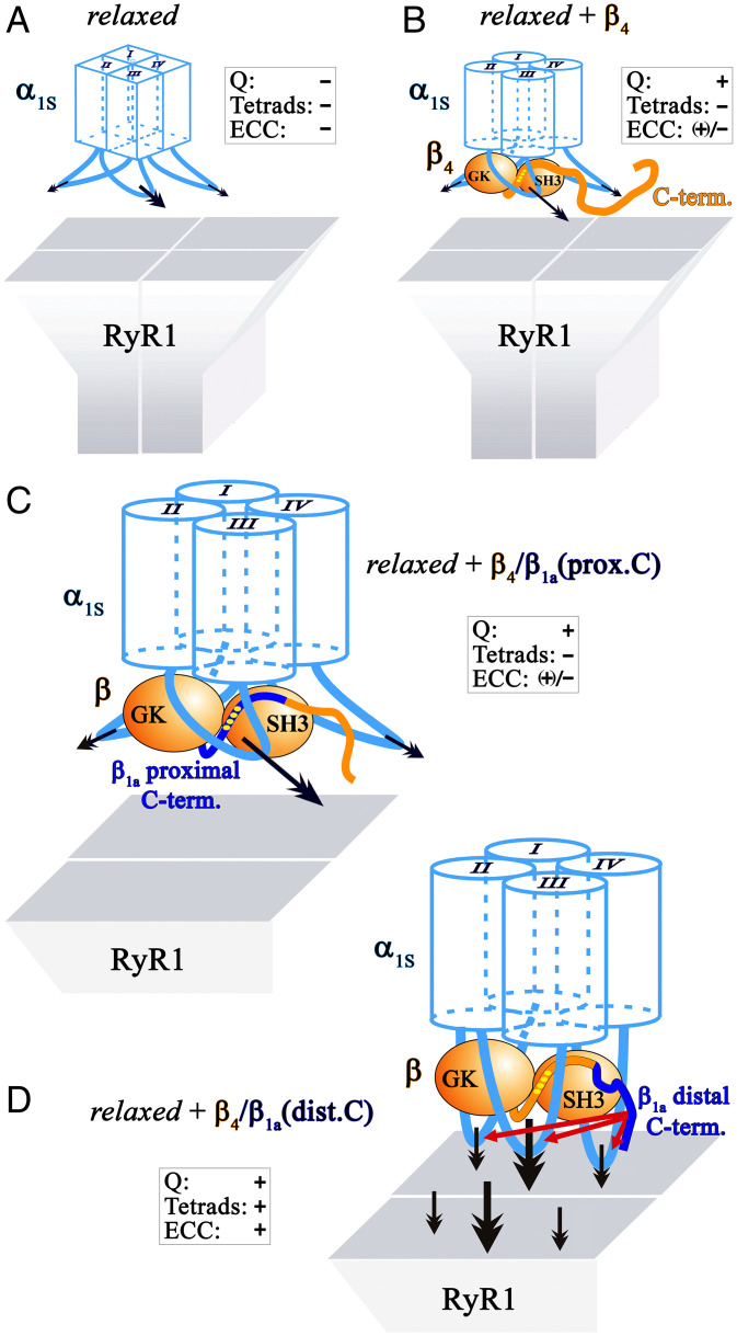
IMAGE

Fig. 7.

ID
ZDB-IMAGE-220717-13
Source
Figures for Dayal et al., 2022
Image
Figure Caption

Fig. 7.

Model of conformational modification of α1S by the β1a distal C terminus—prerequisite for proper skeletal muscle EC coupling. (A) In zebrafish mutant relaxed due to the absence of the DHPRβ1a subunit, the α1S subunit is in a distorted conformation. This causes impediment of charge movement (Q) and of arrangement of DHPR into tetrads (tetrads) that accounts for the lack of skeletal muscle EC coupling (ECC). The distorted conformation of the membrane spanning hydrophobic core regions of the four homologous α1S repeats (I–IV) is depicted by rectangular boxes. The primary and unspecified numbers of secondary α1S-specific RyR1 interaction sites (32) are indicated with bold and normal black arrows, respectively. (B) β4 is unable to reinstate full EC coupling [(+)/−] due to impaired DHPR tetrad formation. According to our model, β4 (symbolized in orange) induces proper conformation of the hydrophobic α1S core regions (depicted with cylinders) required for charge movement function, but is unable to reconstitute accurate conformation of the intracellular α1S loops facilitating RyR1 anchoring (tetrad formation). Improper DHPR–RyR1 interaction (tilted arrows) leads to weak EC coupling and impaired tetrad formation. (C) Likewise, chimera β41a(prox.C) in which the proximal C terminus of β4 is swapped with corresponding β1a sequence (blue), was unable to reinstate intact tetrad formation and thus full ECC. Yellow dots on the proximal C terminus of the β-subunit depict the intramolecular SH3–PXXP interaction sites critical for charge movement function (16). (D) However, the distal C terminus of β1a (blue) enables proper conformation of the intracellular α1S loops crucial for RyR1 anchoring (tetrad formation). Consequently, EC coupling is highly restored upon expression of chimera β41a(dist.C). The direct DHPR–RyR1 interaction depicted in the model is still obscure. However, it is irrelevant for our conclusions whether the two channels interact directly or via an intermediate protein.

Acknowledgments
This image is the copyrighted work of the attributed author or publisher, and ZFIN has permission only to display this image to its users. Additional permissions should be obtained from the applicable author or publisher of the image. Full text @ Proc. Natl. Acad. Sci. USA