FIGURE SUMMARY
Title

Insights into the evolutionary origin of the pineal color discrimination mechanism from the river lamprey

Authors
Wada, S., Kawano-Yamashita, E., Sugihara, T., Tamotsu, S., Koyanagi, M., Terakita, A.
Source
Full text @ BMC Biol.

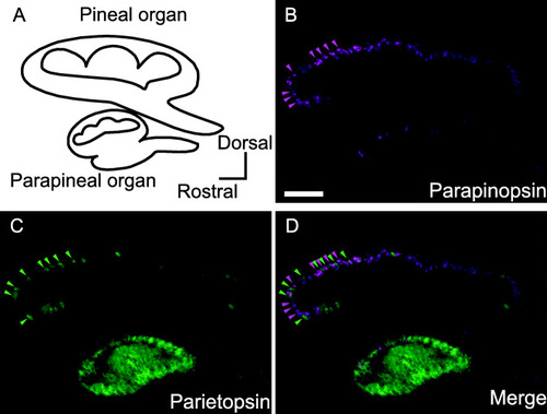
Expression patterns of parapinopsin and parietopsin in the lamprey pineal and parapineal organs. A Lamprey pineal and parapineal organ schematic. BD Fluorescence images by double fluorescence in situ hybridization analyses showing expression patterns of parapinopsin (B, magenta) and parietopsin (C, green). The merged image shows parapinopsin and parietopsin express in separate cells (D). Scale bar = 100 μm

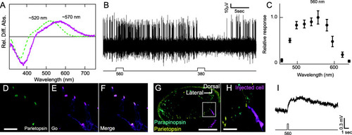
Parietopsin acts as the visible light sensor contributing to pineal color discrimination. A Relative difference absorption spectra of parietopsin reconstituted with 11-cis A1 (green, broken curve) and A2 (magenta, solid curve) retinal before and after light irradiation. Parietopsin with A1 retinal is a green light-sensitive pigment (λmax, ~520 nm) similar to previous reports. Reconstitution with A2 retinal shifts the parietopsin spectrum to longer wavelengths (λmax, ~570 nm). B Chromatic response recorded from pineal ganglion cells showing promotion and inhibition of neural firings to visible light (560 nm) and UV light (380 nm), respectively. C The relative response curve of the visible light-sensitive component (promotion of neural firings) in pineal chromatic responses, showing the response peak at 560 nm, consistent with the previous report [4] (n = 6 measurements, see the “Methods” section and Fig. S3 for the details). Error bars indicate SEM. DF Fluorescence images by immunohistochemical analyses, showing parietopsin (D, green) and Go-type G protein (E, magenta) colocalized in the outer segment (F, merge). Scale bar = 20 μm. GI Analysis of the photoresponse in a parietopsin-expressing photoreceptor cell by intracellular recording. H is the enlarged image from the white square in G. Note that the location of the pineal slice is shown in Fig. S2D. The cell dye loaded by neurobiotin injection (G and H, magenta) expresses parietopsin (yellow) but not parapinopsin (cyan), showing that the intracellular recording was from a single parietopsin-expressing cell. Note that the dye was loaded into the cell body but not into the parietopsin-containing outer segment because the outer segment consists of stacked membranes and limited cytosolic portion like the case of other photoreceptor cells (see Additional file 2: Movie S1). Scale bar = 100 μm (G), 20 μm (H). Intracellular recording analysis of a parietopsin-expressing photoreceptor cell shows a depolarizing response to visible light (I)

Characteristics of parietopsin-expressing photoreceptor cells in the lamprey pineal organ. AC Fluorescence triple staining images showing parietopsin (A, yellow, arrowheads show outer segments), β-arrestin (B, cyan, whole cell), and ganglion cells labeled by retrograde tracing with neurobiotin (B, magenta). The white arrow shows the contact between the parietopsin photoreceptor cells and ganglion cells (C). The dotted trace indicates the outline of the pineal organ. DG Fluorescence quadruple staining images showing localization of β-arrestin (D, white), VGLUT (E, cyan), parietopsin (F, yellow), and parapinopsin (F, magenta). Landmarks with broken line indicate outlines of photoreceptor cells. The yellow and magenta arrowheads in F and G show the outer segments of parietopsin- and parapinopsin-expressing photoreceptor cells, respectively. Both parapinopsin- and parietopsin-expressing photoreceptor cells employ glutamate as a neurotransmitter. Scale bar = 20 μm (A), 10 μm (D)

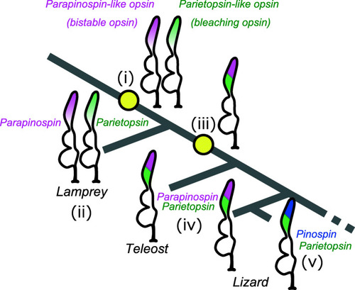
The proposed evolutionary model for color opponency systems in vertebrate pineal-related organs. Bold lines indicate branches of the vertebrate lineage. Speculated ancestral photoreceptor cell types and current types for the pineal color discrimination are drawn on each key node. Please see the “Discussion” section for the detailed descriptions of points (i)–(v)

Acknowledgments
This image is the copyrighted work of the attributed author or publisher, and ZFIN has permission only to display this image to its users. Additional permissions should be obtained from the applicable author or publisher of the image. Full text @ BMC Biol.