
FIGURE
Fig. 1
- ID
- ZDB-FIG-210920-1
- Publication
- Wada et al., 2021 - Insights into the evolutionary origin of the pineal color discrimination mechanism from the river lamprey
- Other Figures
- All Figure Page
- Back to All Figure Page
Fig. 1
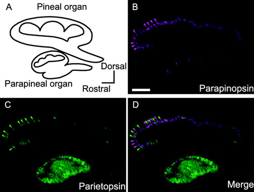
Expression patterns of parapinopsin and parietopsin in the lamprey pineal and parapineal organs. |
Expression Data
Expression Detail
Antibody Labeling
Phenotype Data
Phenotype Detail
Acknowledgments
This image is the copyrighted work of the attributed author or publisher, and
ZFIN has permission only to display this image to its users.
Additional permissions should be obtained from the applicable author or publisher of the image.
Full text @ BMC Biol.