
Fig. 3
- ID
- ZDB-IMAGE-210920-3
- Publication
- Wada et al., 2021 - Insights into the evolutionary origin of the pineal color discrimination mechanism from the river lamprey
- All Figures
- Figures for Wada et al., 2021
|
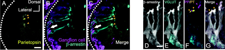
Fig. 3
Characteristics of parietopsin-expressing photoreceptor cells in the lamprey pineal organ.