FIGURE SUMMARY
Title

Rad21l1 cohesin subunit is dispensable for spermatogenesis but not oogenesis in zebrafish

Authors
Blokhina, Y.P., Frees, M.A., Nguyen, A., Sharifi, M., Chu, D.B., Bispo, K., Olaya, I., Draper, B.W., Burgess, S.M.
Source
Full text @ PLoS Genet.

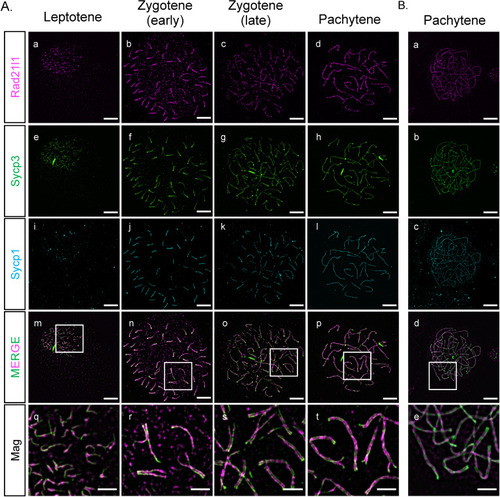
Rad21l1 expression and loading.

(A) Rad21l1 loading during prophase I of meiosis in spermatocyte nuclear surface spreads. Rad21l1 (magenta) loads onto chromosome axes simultaneously with Sycp3 (green) and is also dispersed as foci throughout the spread in leptotene. In early zygotene, Sycp1 (cyan) lines start near the telomeres and synapsis extends inward through late zygotene and are present end-to-end along axes. Note: there is some asynapsis in the pachytene nucleus which may indicate that the cell was either in very early or very late pachytene. The merged images are Rad21l1 and Sycp3 channels only. Mag images are magnifications from the Merge panels; the regions magnified are indicated by white boxes. Panel series a-p scale bar = 5 μm. Mag panel series q-t scale bar = 2 μm. (B) Rad21l1 loading during prophase I of meiosis in oocyte nuclear surface spreads. Panels a-e are arranged similarly to the corresponding panels of part (A).

<italic toggle='yes'>rad21l1</italic><sup><italic toggle='yes'>-/-</italic></sup> mutants are predominantly male due to late sex reversal.

(A) TALEN generated 17-bp deletion leads to a frameshift mutation resulting in a truncated 27 amino acid (aa) Rad21l1 protein with the conserved Rec8/Rad21-like family domains (1–100 aa and 495–543 aa) disrupted or deleted. Rec8/Rad21-like domains (purple boxes); altered amino acid sequence (red box). The ATG translational start site is located at the 4th-6th nucleotide from the end. (B) Spermatocyte nuclear spreads stained for telomeres (cyan), Sycp3 (green), and Rad21l1 (magenta). Rad21l1 forms lines of foci along the Sycp3 axis in rad21l1+/- spermatocytes. In the rad21l1 mutant, no lines of Rad21l1 foci are seen. The rad21l1 mutant spermatocytes can form axes and pair homologs albeit with some asynapsed regions. Scale bar = 5 μm. (C) Sexed offspring of a rad21l1+/- incross show a depletion of females in rad21l1-/- fish. Data pooled from multiple crosses. (D) Sections of gonads prepared from 35–36 dpf rad21l1+/+and rad21l1+/- (labelled rad21l1+) and rad21l1-/- fish and stained for DNA (gray) and Ddx4 (also known as Vasa; green). At 35–36 dpf, oocytes are present in 10/12 rad21l1+/- and 6/11 rad21l1-/- samples. Scale bar = 50 μm. Arrows represent different cell types: (pink- diplotene oocytes, yellow- spermatocytes / sperm; red- premeiotic germ cells). (E) Whole mounts of gonads from 40 and 45 dpf are stained for DNA (gray) and Ddx4 (green). At 40 dpf, oocytes are present in 11/21 rad21l1+/+ and 1/19 rad21l1-/- samples. At 45 dpf, oocytes are present in 7/9 rad21l1+/+ and 1/10 rad21l1-/- samples. Scale bar = 50 μm. Fisher’s exact test used for all statistical analysis. ns = p>0.05, ** = p<0.01, **** = p<0.0001.

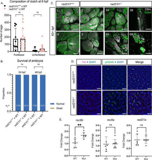
Rad21l1 is dispensable for male fertility.

(A/B) Data resulting from test crosses between rad21l1-/- males and wild-type females to assess fertility and reproductive phenotype. rad21l1+/+ male tank mates were used as controls. No significant difference in the number of eggs the males caused the females to release, the composition of the resulting clutch at 6 hpf, or the survival of the embryos through 48 hpf. Data pooled from 14 crosses over 5 weeks using the same pool of 14 rad21l1-/- males, 12/14 of which crossed successfully at least once. Unpaired, two-tailed student t-test used for statistical analysis, ns = p>0.05. (C) Whole mount adult testes stained for DNA (gray) and Ddx4 (green) showing a phenotypic range of gonad morphology in rad21l1-/- males. All samples except #5 displayed large clusters of mature sperm. Images marked as A and B were taken from the same sample to show variation within a single gonad. Wild-type tank mates used as controls. Scale bar = 50 μm. (D) Testes sections stained with a PNA telomere probe (Tel; magenta), an antibody to γH2AX (green), and DAPI (blue), showing that telomere clustering and DSB localization (γH2AX) are normal in the rad21l1 mutant. Scale bar = 5 μm. (E) Fold change of relative mRNA expression for rad21l1 paralogs, rec8b, rec8a, and rad21a in wild-type versus the rad21l1-/- mutant (Mut). Fold change was determined using the 2-ΔΔCq method. eF1a (housekeeping) mRNA levels were used as a reference.

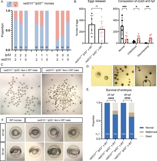
<italic toggle='yes'>tp53</italic> knockout restores females to <italic toggle='yes'>rad21l1</italic> mutant population, but <italic toggle='yes'>rad21l1;tp53</italic> double mutant females produce poor quality eggs and malformed embryos.

(A) Sex ratios of all genotypes resulting from a rad21l1+/-;tp53+/- incross. Data pooled from 3 crosses. 0, 1, and 2 on the x-axis refer to the number of wild-type copies of tp53 and rad21l1. (B) Data resulting from test crosses between rad21l1-/-;tp53-/- females and wild-type males to assess fertility and reproductive phenotype. rad21l1+/+;tp53-/- female tank mates used as controls. No significant difference in the number of eggs the females released. rad21l1-/-;tp53-/- double mutant females release a significantly greater percentage of eggs that fail to be fertilized or display premature decomposition. (C) Representative images of clutches from double mutant and control females at 6 hpf showing lower overall quality of eggs released from double mutant females. (D) Images i-iii show examples of eggs described in the text at 6 hpf. Panel i shows a normal egg (left) and a tiny egg (right). Panel ii shows prematurely decomposing eggs and panel iii shows opaque eggs. (E) Of normal embryos at 6 hpf, 32.4% are dead or malformed at 24 hpf and 38.7% by 48 hpf. Unpaired, two-tailed student t-test used for statistical analysis. (F) Representative images showing the range of malformations seen in developing embryos from rad21l1-/-;tp53-/- females at 24 and 48 hpf. ns = p>0.05, * = p<0.05, ** = p<0.01, *** = p<0.001.

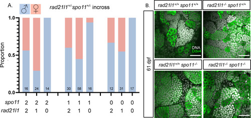
<italic toggle='yes'>spo11;rad21l1</italic> double mutants are infertile males.

(A) Sex ratios of all genotypes resulting from a rad21l1+/-;spo11+/- incross. Data pooled from 6 crosses. 0, 1, and 2 on the x-axis refer to the number of wild-type copies of spo11 and rad21l1. (B) Whole mount testes stained for DNA (gray) and Ddx4 (green). WT and rad21l1-/-;spo11+/+ samples display clusters of mature sperm, while rad21l1+/+;spo11-/- and rad21l1-/-;spo11-/- samples do not. Scale bar = 30 μm.

Acknowledgments
This image is the copyrighted work of the attributed author or publisher, and ZFIN has permission only to display this image to its users. Additional permissions should be obtained from the applicable author or publisher of the image. Full text @ PLoS Genet.