IMAGE

Fig 2

ID
ZDB-IMAGE-210802-26
Source
Figures for Blokhina et al., 2021
Image
Figure Caption

Fig 2 <italic toggle='yes'>rad21l1</italic><sup><italic toggle='yes'>-/-</italic></sup> mutants are predominantly male due to late sex reversal.

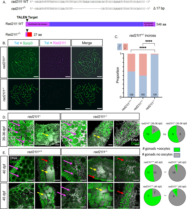
(A) TALEN generated 17-bp deletion leads to a frameshift mutation resulting in a truncated 27 amino acid (aa) Rad21l1 protein with the conserved Rec8/Rad21-like family domains (1–100 aa and 495–543 aa) disrupted or deleted. Rec8/Rad21-like domains (purple boxes); altered amino acid sequence (red box). The ATG translational start site is located at the 4th-6th nucleotide from the end. (B) Spermatocyte nuclear spreads stained for telomeres (cyan), Sycp3 (green), and Rad21l1 (magenta). Rad21l1 forms lines of foci along the Sycp3 axis in rad21l1+/- spermatocytes. In the rad21l1 mutant, no lines of Rad21l1 foci are seen. The rad21l1 mutant spermatocytes can form axes and pair homologs albeit with some asynapsed regions. Scale bar = 5 μm. (C) Sexed offspring of a rad21l1+/- incross show a depletion of females in rad21l1-/- fish. Data pooled from multiple crosses. (D) Sections of gonads prepared from 35–36 dpf rad21l1+/+and rad21l1+/- (labelled rad21l1+) and rad21l1-/- fish and stained for DNA (gray) and Ddx4 (also known as Vasa; green). At 35–36 dpf, oocytes are present in 10/12 rad21l1+/- and 6/11 rad21l1-/- samples. Scale bar = 50 μm. Arrows represent different cell types: (pink- diplotene oocytes, yellow- spermatocytes / sperm; red- premeiotic germ cells). (E) Whole mounts of gonads from 40 and 45 dpf are stained for DNA (gray) and Ddx4 (green). At 40 dpf, oocytes are present in 11/21 rad21l1+/+ and 1/19 rad21l1-/- samples. At 45 dpf, oocytes are present in 7/9 rad21l1+/+ and 1/10 rad21l1-/- samples. Scale bar = 50 μm. Fisher’s exact test used for all statistical analysis. ns = p>0.05, ** = p<0.01, **** = p<0.0001.

Acknowledgments
This image is the copyrighted work of the attributed author or publisher, and ZFIN has permission only to display this image to its users. Additional permissions should be obtained from the applicable author or publisher of the image. Full text @ PLoS Genet.