FIGURE SUMMARY
Title

Social Experience Regulates Endocannabinoids Modulation of Zebrafish Motor Behaviors

Authors
Orr, S.A., Ahn, S., Park, C., Miller, T.H., Kassai, M., Issa, F.A.
Source
Full text @ Front. Behav. Neurosci.

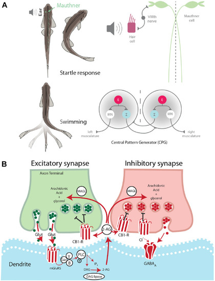
Startle escape and swim motor behaviors are socially regulated. (A) Startle behavior in zebrafish is controlled by the M-cell startle escape circuit. The auditory startle escape response is activated when a sound activates hair cells within the ear. Next, the signal is sent from the VIIIth nerve to the M-cell, which activates contralateral fast motor neurons responsible for contraction of flexor muscles that leads to the startle escape response. The swimming motor pattern is controlled by the central pattern generators (CPGs) which repeat along the length of the spinal cord. Each half-center of the CPG is composed of an excitatory interneuron (E), an inhibitory interneuron (I), and a motor neuron (MN). The motor neurons project ipsilaterally to the trunk musculature and induce contraction. (B) Schematic model of endocannabinoid retrograde signaling. The endocannabinoid 2-AG is synthesized post-synaptically in response to neurotransmitter binding. Traditional model suggests that retrograde transmission of 2-AG inhibits further release from both excitatory and inhibitory neurons. DAG lipase synthesizes 2-AG post-synaptically. CB1 receptor binds 2-AG. MAGL degrades 2-AG in presynaptic terminal.

Endocannabinoid signaling pathway is socially regulated and its modulation of M-cell excitability is status-dependent. (A) qPCR gene expression analysis of ECS signaling molecules. Data of dominants and subordinates normalized to control isolates (n = 6 pairs; control isolates n = 6; Kruskal–Wallis test, *p < 0.05). (B) Western blot analysis of dagl-α and cb1 receptor. Each protein was tested concurrently with β-actin as a control. Protein expression of dominants and subordinates was normalized to communal controls as a ratio (illustrated values below each band); C, communals; D, dominants; S, subordinates. Bar graphs represent average % change in protein concentration of four replicates of samples each consisting of 10 brains normalized to WT communals.

2-AG modulation of escape and swimming activities is social status-dependent. (A–C) Probability of startle escape response before (control) and after JZL184 injections for communals, dominants and subordinates, respectively. Asterisks (p < 0.05, ∗∗p < 0.005) denote statistical difference between control and experimental condition at the specified dB level. (D) 1 min recoding of far field-potentials of spontaneous swimming activity before (control) and (E) after JZL184 injections for communal, dominants and subordinates, respectively, along with respective raster plots of each condition. (F) Average swimming frequency for all animals tested before and after JZL184 injection. (G) Box and whiskers plots of the average number of swim bursts per 1 min for each social phenotype. Dots represent individual animals. The box extends from the 25th to 75th percentiles, horizontal line is the median, and whiskers represent max/min values.

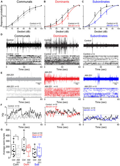
Effect of AM-251 on status-dependent escape probability and swim frequency. (A–C) Probability of startle escape response before (control) and after AM-251 injections for communals, dominants and subordinates, respectively. Asterisks (*p < 0.05) denote statistical difference between control and experimental condition at the specified dB level. (D,E) 1 min recoding of far field-potentials of spontaneous swimming activity before (control) and after AM-251 injections for communal, dominants and subordinates, respectively, along with respective raster plots for all animals tested. (F) Average swimming frequency for all animals tested before and after AM-251 injection. (G) Box and whiskers plots of the average number of swim bursts per 1 min for each social phenotype. Box plot parameters are defined in Figure 3G.

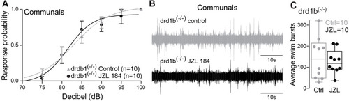
drd1b expression is socially regulated and necessary for status-dependent ECS regulation of escape and swim circuits. (A) Probability of startle escape response of drd1b(–/–) communal zebrafish before (control) and after injection of JZL184. (B) 1 min recording of far field-potentials of spontaneous swimming activity before (control) and after JZL184 injections for drd1b(–/–) communal zebrafish. (C) Average number of swim bursts per 1 min for drd1b(–/–) communal fish (n = 10) before and after JZL184 injection.

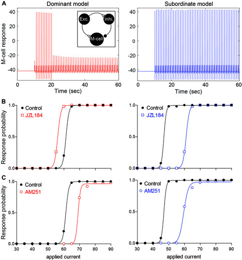
Neurocomputational models simulating the effects of 2-AG on the escape response. (A) Examples of the response of the M-cell model cell within a simple model network (inset) to repeated suprathreshold applied current injection in dominant-like (left) and subordinate-like (right) models. (B,C) Results of dominant-like and subordinate-like models simulating the probability of startle escape response before (control, black solid line) and during [JZL184, (B)] and blockage of CB1R [AM-251, (C)].

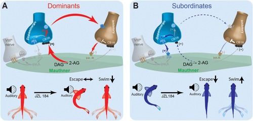
Schematic model for social status-dependent regulation of neurochemical inputs to the M-cell: The M-cell (green) receives inputs from DA cells (blue), the excitatory VIIIth cranial nerve (gray), and inhibitory (brown). Our model predicts distinct neurochemical pathways in dominants (A) and subordinates (B) responsible for differences in startle escape sensitivity. These pathways are proposed based on differential effects of JZL184 treatment on startle escape behavior (bottom). Higher baseline 2-AG in dominants is responsible for activation of the “inhibitory pathway” via inhibitory neurotransmitter release. Lower baseline 2-AG in subordinates activates the lower threshold “excitatory pathway” via the VIIIth nerve, responsible for higher startle escape sensitivity.

Acknowledgments
This image is the copyrighted work of the attributed author or publisher, and ZFIN has permission only to display this image to its users. Additional permissions should be obtained from the applicable author or publisher of the image. Full text @ Front. Behav. Neurosci.