FIGURE SUMMARY
Title

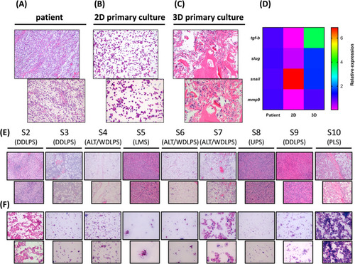
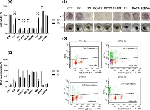
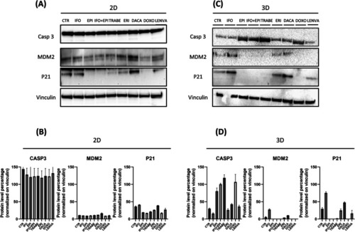
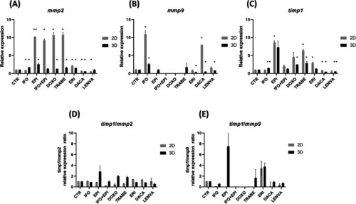
The potential role of the extracellular matrix in the activity of trabectedin in UPS and L-sarcoma: evidences from a patient-derived primary culture case series in tridimensional and zebrafish models

Authors
De Vita, A., Recine, F., Miserocchi, G., Pieri, F., Spadazzi, C., Cocchi, C., Vanni, S., Liverani, C., Farnedi, A., Fabbri, F., Fausti, V., Casadei, R., Brandolini, F., Ercolani, G., Cavaliere, D., Bongiovanni, A., Riva, N., Gurrieri, L., Di Menna, G., Calpona, S., Debonis, S.A., Mercatali, L., Ibrahim, T.
Source
Full text @ J. Exp. Clin. Cancer Res.

a hematoxylin and eosin staining of the patient surgical specimen showing high-grade polymorphic UPS cells (light blue stroma) infiltrating necrotic tissue, 10 x and 20 x magnification. b hematoxylin and eosin staining of the cytospunned tumor cells from the patient-derived UPS primary culture S1, 10 x and 20 x magnification. c hematoxylin and eosin staining of the cytospunned tumor cells (light blue spots) from the patient-derived UPS primary culture S1 cultured within 3D collagen-based scaffold system, 10 x and 20 x magnification. d Heat map comparisons of the relative gene expression of selected tumor-associated markers tgf-b, slug, snail, mmp9 between UPS patient surgical specimen, S1 patient-derived 2D standard monolayer primary culture and S1 patient-derived 3D primary culture system. e hematoxylin and eosin staining of the patient surgical specimen showing low and high grade UPS and L-sarcoma (light blue stroma) infiltrating adipose tissue, 10x and 20× magnification. f hematoxylin and eosin staining of the cytospunned tumor cells of patient-derived primary cultures UPS and L-sarcoma case series, 10 x and 20 x magnification

a Chemobiogram analysis of S1 primary culture seeded in 2D and 3D-collagen based scaffold and exposed to chemotherapeutics agent, untreated cells were used as control. b Representative images of 2D and 3D-collagen based scaffold S1 primary culture exposed to the tested drugs. c DNA fragmentation analysis expressed as apoptotic cells % of 2D and 3D-collagen based scaffold S1 primary culture exposed to the tested drugs. d Representative images (dot plot) of DNA fragmentation analysis obtained thought flow cytometry

a Proteomic analysis of apoptotic- and anti-apoptotic-related proteins in 2D S1 primary culture exposed to the tested drugs. b Densitometric analysis of protein bands % normalized on the housekeeping vinculin. c Proteomic analysis of apoptotic- and anti-apoptotic-related proteins in 3D-collagen based scaffold S1 primary culture exposed to the tested drugs. d Densitometric analysis of protein bands % normalized on the housekeeping vinculin

a-c Relative expression of ECM-associated genes in 2D and 3D-collagen based scaffold S1 primary culture. d timp1/mmp2 relative expression ratio genes in 2D and 3D-collagen based scaffold S1 primary culture. e timp1/mmp9 relative expression ratio genes in 2D and 3D-collagen based scaffold S1 primary culture

a Representative fluorescence microscopy images of zebrafish embryos xenotrasnplanted with S1 cultured in standard monolayer culture (2D) and within 3D collagen-based scaffold (3D). Images of embryos untreated at 2 and 72 h post injection and exposed to trabectedin at 72 h post injection, scale bar 1000 μm. b Mean fluorescence signal of 2D and 3D xenotransplanted embryos, arbitrary units. c Tumor growth inhibition rate between 2D and 3D groups

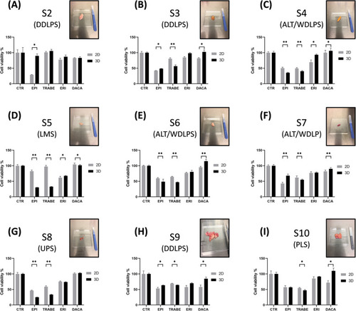
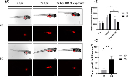
Pharmacological profile of 2D and 3D-collagen based scaffold UPS and L-sarcoma primary culture case series. Primary cells were exposed to selected first- and second- line treatments (EPI, TRABE, ERI, DACA) for STS. Images of the surgical specimens used for the establishment of primary cultures are reported (a) S2 DDLPS. b S3 DDLPS. c S4 ALT/WDLPS. d S5 LMS. e S6 ALT/WDLPS. f S7 ALT/WDLPS. g S8 UPS. h DDLPS. i PLS

Acknowledgments
This image is the copyrighted work of the attributed author or publisher, and ZFIN has permission only to display this image to its users. Additional permissions should be obtained from the applicable author or publisher of the image. Full text @ J. Exp. Clin. Cancer Res.