IMAGE

Fig. 1

ID
ZDB-IMAGE-210518-27
Source
Figures for De Vita et al., 2021
Image
Figure Caption

Fig. 1

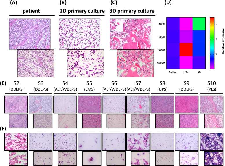
a hematoxylin and eosin staining of the patient surgical specimen showing high-grade polymorphic UPS cells (light blue stroma) infiltrating necrotic tissue, 10 x and 20 x magnification. b hematoxylin and eosin staining of the cytospunned tumor cells from the patient-derived UPS primary culture S1, 10 x and 20 x magnification. c hematoxylin and eosin staining of the cytospunned tumor cells (light blue spots) from the patient-derived UPS primary culture S1 cultured within 3D collagen-based scaffold system, 10 x and 20 x magnification. d Heat map comparisons of the relative gene expression of selected tumor-associated markers tgf-b, slug, snail, mmp9 between UPS patient surgical specimen, S1 patient-derived 2D standard monolayer primary culture and S1 patient-derived 3D primary culture system. e hematoxylin and eosin staining of the patient surgical specimen showing low and high grade UPS and L-sarcoma (light blue stroma) infiltrating adipose tissue, 10x and 20× magnification. f hematoxylin and eosin staining of the cytospunned tumor cells of patient-derived primary cultures UPS and L-sarcoma case series, 10 x and 20 x magnification

Acknowledgments
This image is the copyrighted work of the attributed author or publisher, and ZFIN has permission only to display this image to its users. Additional permissions should be obtained from the applicable author or publisher of the image. Full text @ J. Exp. Clin. Cancer Res.