IMAGE

Fig. 5

ID
ZDB-IMAGE-210518-13
Source
Figures for De Vita et al., 2021
Image
Figure Caption

Fig. 5

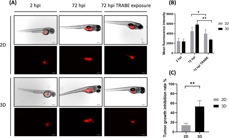
a Representative fluorescence microscopy images of zebrafish embryos xenotrasnplanted with S1 cultured in standard monolayer culture (2D) and within 3D collagen-based scaffold (3D). Images of embryos untreated at 2 and 72 h post injection and exposed to trabectedin at 72 h post injection, scale bar 1000 μm. b Mean fluorescence signal of 2D and 3D xenotransplanted embryos, arbitrary units. c Tumor growth inhibition rate between 2D and 3D groups

Acknowledgments
This image is the copyrighted work of the attributed author or publisher, and ZFIN has permission only to display this image to its users. Additional permissions should be obtained from the applicable author or publisher of the image. Full text @ J. Exp. Clin. Cancer Res.