
- Title
-
A Novel Infection Protocol in Zebrafish Embryo to Assess Pseudomonas aeruginosa Virulence and Validate Efficacy of a Quorum Sensing Inhibitor In Vivo
- Authors
- Nogaret, P., El Garah, F., Blanc-Potard, A.B.
- Source
- Full text @ Pathogens

Development of bath infection model with injured zebrafish embryos to assess P. aeruginosa virulence. (A) Larvae were injured in the tail fin at 48 hours post-fertilization (hpf). (B) Survival curves (Kaplan–Meier representation) of healthy or injured embryos (GAB line) immersed at 48 hpf with PAO1 wild-type strain at approximately 2*107 CFU/mL grown in exponential phase or “fish water” (negative control). (C) Survival curves (Kaplan–Meier representation) of injured embryos (GAB line) immersed at 48 hpf with PAO1 strain at two different concentrations (3.5 × 10⁷ or 7 × 10⁶ CFU/mL) or fish water (negative control). Results are expressed as the percentage of surviving embryos. Pools of three biologically independent replicates are shown for each survival curve (in each experiment 20 embryos were used per strain). Significant difference at ** p < 0.01, *** p < 0.001 or no significant difference: ns. (D) In vivo imaging of injured embryos immersed with fluorescent P. aeruginosa PAO1-GFP for 1.5 or 24 hours (left panels). Images represent DIC channel merge to green fluorescence protein (GFP) channel. Scale bar: 0.5 mm. Images of injured embryos immersed with N-phenylthiourea (PTU) without bacteria (right panels) are shown as control. Images are a representative result of three embryos in each condition. |
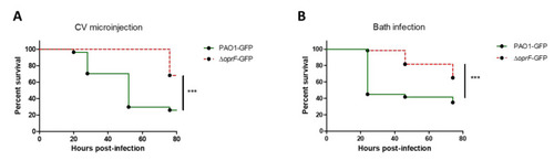
Validation of bath infection model using an attenuated strain of P. aeruginosa. (A) Kaplan–Meier representation of survival after microinjection of approximately 3500 CFUs of wild-type or ΔoprF GFP-expressing P. aeruginosa into the caudal vein (CV) of embryos. Pools of three biologically independent replicates are shown (in each experiment 20 embryos were used per strain). (B) Kaplan–Meier representation of survival after bath infection of injured embryos with wild-type or ΔoprF GFP-expressing P. aeruginosa at approximately 108 CFU/mL. Pools of three biologically independent replicates are shown for each survival curve (in each experiment 20 embryos were used per strain). |
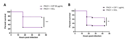
Validation of bath infection model with a known antibiotic. (A) Kaplan–Meier representation of the survival of injured zebrafish embryos bath infected with PAO1 strain at approximately 108 CFU/mL and treated with ciprofloxacin 50 µg/mL or hydrochloric acid 0.2 mM (negative control) 2 hours after infection. (B) Kaplan–Meier representation of the survival of injured zebrafish embryos bath infected with PAO1 strain at approximately 5 × 10⁷ CFU/mL and treated with ciprofloxacin 1 µg/mL or hydrochloric acid 0.5 mM (negative control) 2 hours after infection. Pools of three biologically independent replicates (with 20 embryos) are shown for each survival curve. *** p < 0.001; ** p < 0.01. |

Use of bath infection model to validate novel anti-Pseudomonas molecules such as C11 molecule (targeting quorum sensing). (A) Schematic protocol for testing antivirulence molecules in embryos bath infected with P. aeruginosa. Antivirulence molecules, which do not reduce bacterial viability outside the host, are added together with bacteria. (B) The antivirulence efficacy of C11 (dissolved in DMSO) was tested with embryos injured in the tail fin and bath infected with PAO1 suspension at approximately 107 CFU/mL in presence of C11 at 10, 25, or 50 µM. Injured embryos in the control group were treated with PAO1 suspension in presence of DMSO at 0.05, 0.13, or 0.25% (reflecting the amount of DMSO in the C11-treated groups). A significant difference (** p < 0.05, *** p < 0.001) is found in the survival curve of C11-treated embryos compared to non-treated embryos at 25 or 50 µM. (C) The toxicity of C11 at 25 µM was monitored after immersion of embryos injured in the tail fin. For all experiments, the embryo survival was monitored for 45 hours and survival curves were represented with a Kaplan–Meier representation. Graphs represent the pool of three independent experiments (n = 60 larvae in total per condition). |