
|
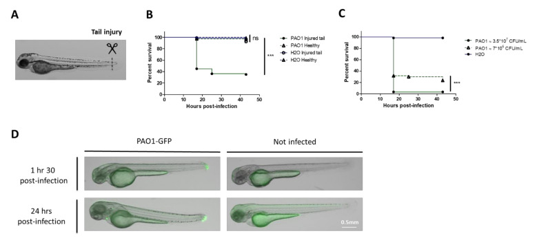
Figure 1 Development of bath infection model with injured zebrafish embryos to assess P. aeruginosa virulence. (A) Larvae were injured in the tail fin at 48 hours post-fertilization (hpf). (B) Survival curves (Kaplan–Meier representation) of healthy or injured embryos (GAB line) immersed at 48 hpf with PAO1 wild-type strain at approximately 2*107 CFU/mL grown in exponential phase or “fish water” (negative control). (C) Survival curves (Kaplan–Meier representation) of injured embryos (GAB line) immersed at 48 hpf with PAO1 strain at two different concentrations (3.5 × 10⁷ or 7 × 10⁶ CFU/mL) or fish water (negative control). Results are expressed as the percentage of surviving embryos. Pools of three biologically independent replicates are shown for each survival curve (in each experiment 20 embryos were used per strain). Significant difference at ** p < 0.01, *** p < 0.001 or no significant difference: ns. (D) In vivo imaging of injured embryos immersed with fluorescent P. aeruginosa PAO1-GFP for 1.5 or 24 hours (left panels). Images represent DIC channel merge to green fluorescence protein (GFP) channel. Scale bar: 0.5 mm. Images of injured embryos immersed with N-phenylthiourea (PTU) without bacteria (right panels) are shown as control. Images are a representative result of three embryos in each condition.