
- Title
-
Development of the CK-MB-1 trastuzumab-resistant HER2-positive breast cancer cell line and xenograft animal models
- Authors
- Chung, W.P., Huang, W.L., Liao, W.A., Huang, W.L., Liu, Y.Y., Su, W.C.
- Source
- Full text @ Cancer Med

Summary of patient treatment. Before the development of malignant ascites, the patient received three lines of anti‐HER2 treatment for metastatic disease and following disease progression. FEC, fluorouracil, epirubicin, and cyclophosphamide; TH, docetaxel and trastuzumab; PD, progressive disease |

Characteristics of the CK‐MB‐1 cell line. (A) Western blotting confirmed HER2 positivity, ER/PR negativity, and no expression of EGFR in CK‐MB‐1 cells. (B) There was no loss of PTEN protein expression in CK‐MB‐1 cells. (C) HER2 gene amplification was detected by FISH. Orange signals were HER2 genes and green signals were chromosome 17 centromeres. (D) Sanger sequencing of CK‐MB‐1 cells did not reveal common |
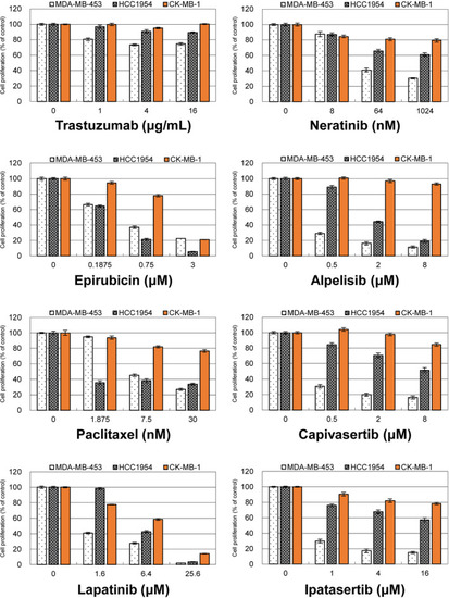
Antiproliferative effects of different drugs in three trastuzumab‐resistant cell lines. MDA‐MB‐453, HCC1954, and CK‐MB‐1 cells were treated with one anti‐HER2 monoclonal antibody, two chemotherapies, and five tyrosine kinase inhibitors targeting HER2, PI3K, or AKT. These drugs were less effective against the proliferation of CK‐MB‐1 cells than against the other two trastuzumab‐resistant cell lines. HER2, human epidermal growth factor receptor 2; PI3K, phosphoinositide‐3‐kinase |
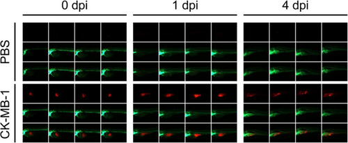
The zebrafish xenograft model of the CK‐MB‐1 cell line. Tg(fli1: EGFP) transgenic zebrafish were microinjected with PBS and CK‐MB‐1 cells ( |
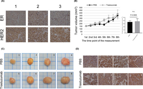
Xenograft mouse models of the CK‐MB‐1 cell line. (A) IHC staining revealed that CK‐MB‐1 xenografts maintained the same ER‐negative/HER2‐positive profile of the parental cell line in the ASID mouse model. (B) NOD‐SCID mice carrying CK‐MB‐1 xenografts were intraperitoneally injected with PBS or trastuzumab ( |