
|
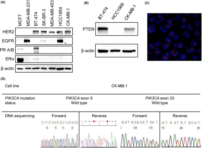
FIGURE 2
Characteristics of the CK‐MB‐1 cell line. (A) Western blotting confirmed HER2 positivity, ER/PR negativity, and no expression of EGFR in CK‐MB‐1 cells. (B) There was no loss of PTEN protein expression in CK‐MB‐1 cells. (C) HER2 gene amplification was detected by FISH. Orange signals were HER2 genes and green signals were chromosome 17 centromeres. (D) Sanger sequencing of CK‐MB‐1 cells did not reveal common